Application
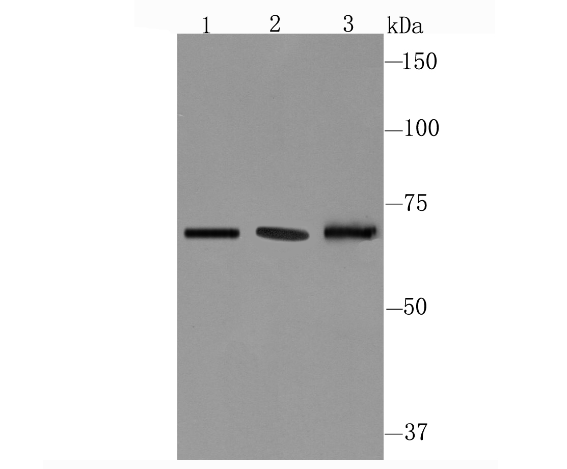
Fig1: Western blot analysis of CD39 on different cell lysate using anti-CD39 antibody at 1/1,000 dilution. Positive control: Lane1: Human spleen Lane2: Mouse placenta Lane3: Mouse spleenApplication
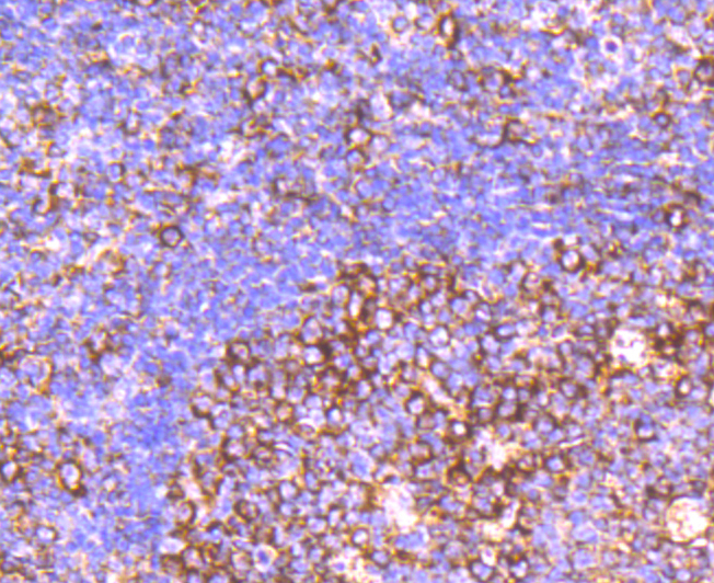
Fig2: Immunohistochemical analysis of paraffin-embedded human tonsil tissue using anti-CD39 antibody. Counter stained with hematoxylin.Application
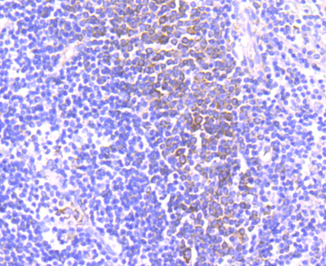
Fig3: Immunohistochemical analysis of paraffin-embedded human spleen tissue using anti-CD39 antibody. Counter stained with hematoxylin.Application
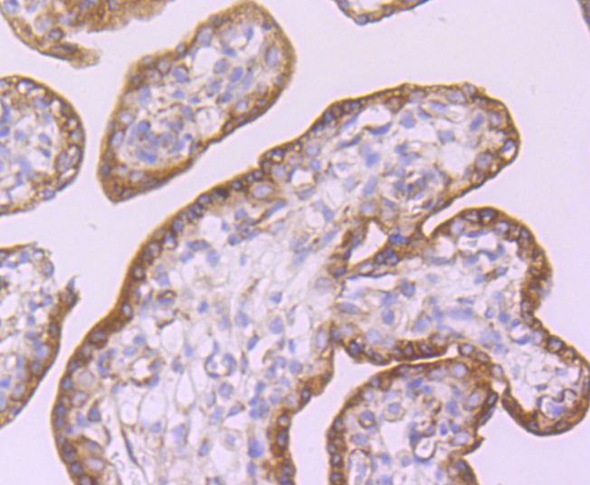
Fig4: Immunohistochemical analysis of paraffin-embedded human placenta tissue using anti-CD39 antibody. Counter stained with hematoxylin.Application
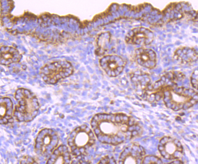
Fig5: Immunohistochemical analysis of paraffin-embedded mouse uterus tissue using anti-CD39 antibody. Counter stained with hematoxylin.| Product Name | CD39 [JA90-36] |
|---|---|
| Antibody Type | Primary Antibodies |
| Antigen Alias | ATPDase antibody CD 39 antibody CD39 antibody CD39 antigen antibody DKFZp686D194 antibody DKFZp686I093 antibody Ecto apyrase antibody Ecto ATP diphosphohydrolase antibody Ecto-apyrase antibody Ecto-ATP diphosphohydrolase 1 antibody Ecto-ATPase 1 antibody Ecto-ATPDase 1 antibody Ectonucleoside triphosphate diphosphohydrolase 1 antibody ENTP1_HUMAN antibody ENTPD 1 antibody ENTPD1 antibody FLJ40921 antibody FLJ40959 antibody Lymphoid cell activation antigen antibody NTPDase 1 antibody NTPDase1 antibody SPG64 antibody |
| Product description | CD39, also known as ectonucleoside triphosphate diphosphohydrolase 1 (ENP1), is an integral membrane glycoprotein that acts as an extracellular nucleotide-hydrolyzing enzyme. CD39 inhibits ADP-induced platelet aggregation by hydrolyzing ADP to AMP and ultimately generating adenosine. Intracellular CD39 undergoes glycosylation at 6 N-glycosylation sites and translocates to the membrane in order to be an active enzyme. Alternative splicing gives rise to three CD39 isoforms, vascular, placenta I and placenta II. The placenta I isoform differs at the amino terminus whereas the placenta II isoform is missing amino acids 300-510 at the C-terminus. CD39 is expressed in vascular tissues including placenta, lung, skeletal muscle and kidney, as well as endothelium, smooth muscle, cardiac cells, lymphocytes (such as activated B cells) activated NK cells, macrophages, dendridic cells and platelets. CD39 may be used as an anti-thrombic agent for pre-treating patients at risk for coronary artery occlusion and thrombic stroke. |
| Immunogen | recombinant protein |
| Clonality | Monoclonal |
|---|---|
| Isotype | IgG |
| Host Species | Recombinant rabbit |
| Tested Applications | WBIHC |
| WB:1:500-1:1,000 IHC:1:50-1:200 |
|
| Species Reactivity | HumanMouseRat |
| Concentration | 1mg/ml |
| Alternative Names | ATPDase antibody CD 39 antibody CD39 antibody CD39 antigen antibody DKFZp686D194 antibody DKFZp686I093 antibody Ecto apyrase antibody Ecto ATP diphosphohydrolase antibody Ecto-apyrase antibody Ecto-ATP diphosphohydrolase 1 antibody Ecto-ATPase 1 antibody Ecto-ATPDase 1 antibody Ectonucleoside triphosphate diphosphohydrolase 1 antibody ENTP1_HUMAN antibody ENTPD 1 antibody ENTPD1 antibody FLJ40921 antibody FLJ40959 antibody Lymphoid cell activation antigen antibody NTPDase 1 antibody NTPDase1 antibody SPG64 antibody |
|---|---|
| Molecular Weight(MW) | 70 kDa |
| Cellular Localization | Membrane. |
Application
Fig1: Western blot analysis of CD39 on different cell lysate using anti-CD39 antibody at 1/1,000 dilution. Positive control: Lane1: Human spleen Lane2: Mouse placenta Lane3: Mouse spleen
Application
Fig2: Immunohistochemical analysis of paraffin-embedded human tonsil tissue using anti-CD39 antibody. Counter stained with hematoxylin.
Application
Fig3: Immunohistochemical analysis of paraffin-embedded human spleen tissue using anti-CD39 antibody. Counter stained with hematoxylin.
Application
Fig4: Immunohistochemical analysis of paraffin-embedded human placenta tissue using anti-CD39 antibody. Counter stained with hematoxylin.
Application
Fig5: Immunohistochemical analysis of paraffin-embedded mouse uterus tissue using anti-CD39 antibody. Counter stained with hematoxylin.| Positive Control | Human spleen tissue lysate, mouse placenta tissue, mouse spleen tissue, human tonsil tissue, human spleen tissue, human placenta tissue, mouse uterus tissue. |
|---|---|
| Application Notes | WB:1:500-1:1,000 IHC:1:50-1:200 |
| Form | Liquid |
|---|---|
| Storage Instructions | Store at +4℃ after thawing. Aliquot store at -20℃ or -80℃. Avoid repeated freeze / thaw cycles. |
| Storage Buffer | 1*TBS (pH7.4), 1%BSA, 40%Glycerol. Preservative: 0.05% Sodium Azide. |


