Application
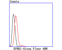
Fig4: Flow cytometric analysis of Raji cells with SPHK1 antibody at 1/100 dilution (red) compared with an unlabelled control (cells without incubation with primary antibody; black).Application
Fig4: Flow cytometric analysis of Raji cells with SPHK1 antibody at 1/100 dilution (red) compared with an unlabelled control (cells without incubation with primary antibody; black).Application
Fig4: Flow cytometric analysis of Raji cells with SPHK1 antibody at 1/100 dilution (red) compared with an unlabelled control (cells without incubation with primary antibody; black).Application
Fig4: Flow cytometric analysis of Raji cells with SPHK1 antibody at 1/100 dilution (red) compared with an unlabelled control (cells without incubation with primary antibody; black).| Product Name | SPHK1 [JA31-14] |
|---|---|
| Antibody Type | Primary Antibodies |
| Antigen Alias | SK 1 antibody SK1 antibody Sphingosine kinase 1 antibody SPHK 1 antibody SPHK antibody Sphk1 antibody SPHK1_HUMAN antibody SPK antibody SPK 1 antibody |
| Product description | Sphingosine kinase (SphK or SphK1) is a key enzyme catalyzing the phosphorylation of sphingosine to form sphingosine 1-phosphate (SPP or S1P). Competitive inhibitors of SphK1 block formation of SPP and selectively inhibit cellular proliferation induced by a variety of factors. One potent inhibitor of SphK1 activity is DMS (N,N-dimethylsphingosine). SPP/SphK1 has been implicated as a signaling pathway that regulates diverse cellular functions, including cell growth, proliferation and survival. Specifically, SphK1 is involved in the signaling pathway(s) that protects human hepatocytes from the apoptotic action of TNFα. Furthermore, SPP/SphK1 may play an important role in neuronal survival by regulating activation of SAPKs and caspases. SphK1 is widely expressed with highest levels in adult liver, kidney, heart and skeletal muscle; however, activation of SphK1 disengages cells from their liver-specific phenotype. |
| Immunogen | recombinant protein |
| Clonality | Monoclonal |
|---|---|
| Isotype | IgG |
| Host Species | Recombinant rabbit |
| Tested Applications | WBIHCFC |
| WB:1:500-1:1,000 IHC:1:50-1:200 FC:1:50 |
|
| Species Reactivity | HumanMouseRat |
| Concentration | 1mg/ml |
| Alternative Names | SK 1 antibody SK1 antibody Sphingosine kinase 1 antibody SPHK 1 antibody SPHK antibody Sphk1 antibody SPHK1_HUMAN antibody SPK antibody SPK 1 antibody |
|---|---|
| Molecular Weight(MW) | 51 kDa |
| Cellular Localization | Cytoplasm. Nucleus. Membrane. |
Application
Fig4: Flow cytometric analysis of Raji cells with SPHK1 antibody at 1/100 dilution (red) compared with an unlabelled control (cells without incubation with primary antibody; black).
Application
Fig4: Flow cytometric analysis of Raji cells with SPHK1 antibody at 1/100 dilution (red) compared with an unlabelled control (cells without incubation with primary antibody; black).
Application
Fig4: Flow cytometric analysis of Raji cells with SPHK1 antibody at 1/100 dilution (red) compared with an unlabelled control (cells without incubation with primary antibody; black).
Application
Fig4: Flow cytometric analysis of Raji cells with SPHK1 antibody at 1/100 dilution (red) compared with an unlabelled control (cells without incubation with primary antibody; black).| Positive Control | C2C12, Raji, human kidney tissue, rat kidney tissue. |
|---|---|
| Application Notes | WB:1:500-1:1,000 IHC:1:50-1:200 FC:1:50 |
| Form | Liquid |
|---|---|
| Storage Instructions | Store at +4℃ after thawing. Aliquot store at -20℃ or -80℃. Avoid repeated freeze / thaw cycles. |
| Storage Buffer | 1*TBS (pH7.4), 1%BSA, 40%Glycerol. Preservative: 0.05% Sodium Azide. |


