Application
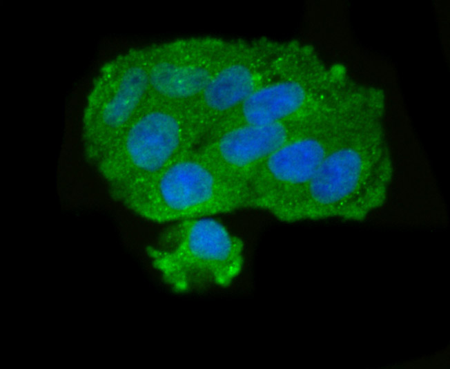
Fig1: ICC staining GOLPH3 in Hela cells (green). The nuclear counter stain is DAPI (blue). Cells were fixed in paraformaldehyde, permeabilised with 0.25% Triton X100/PBS.Application
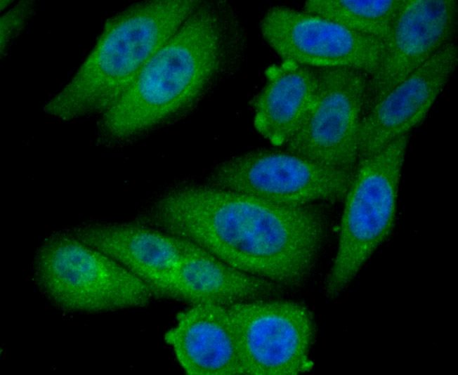
Fig2: ICC staining GOLPH3 in HepG2 cells (green). The nuclear counter stain is DAPI (blue). Cells were fixed in paraformaldehyde, permeabilised with 0.25% Triton X100/PBS.Application
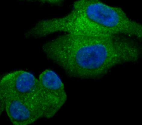
Fig3: ICC staining GOLPH3 in SK-Br-3 cells (green). The nuclear counter stain is DAPI (blue). Cells were fixed in paraformaldehyde, permeabilised with 0.25% Triton X100/PBS.Application
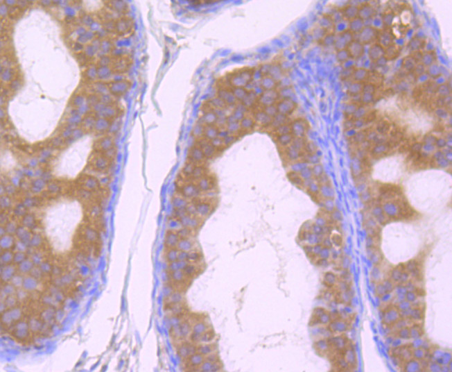
Fig4: Immunohistochemical analysis of paraffin-embedded rat epididymis tissue using anti-GOLPH3 antibody. Counter stained with hematoxylin.Application
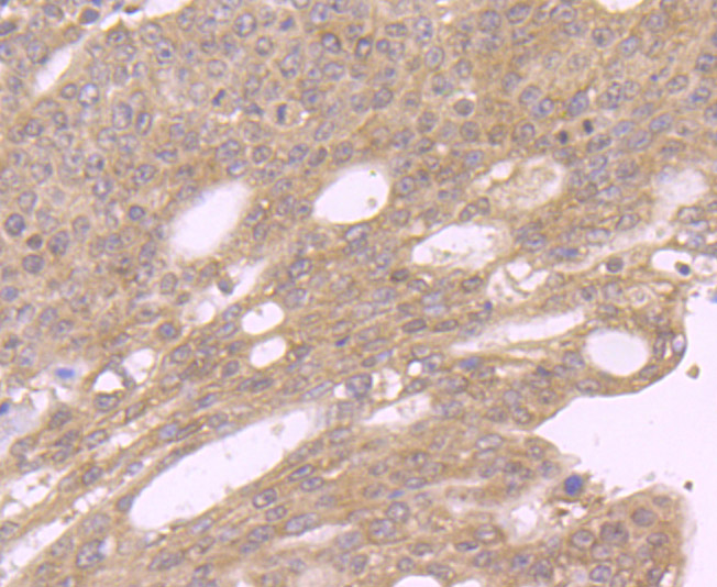
Fig5: Immunohistochemical analysis of paraffin-embedded human stomach cancer tissue using anti-GOLPH3 antibody. Counter stained with hematoxylin.Application
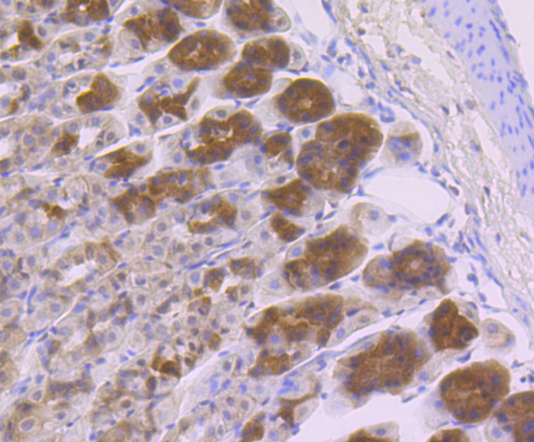
Fig6: Immunohistochemical analysis of paraffin-embedded mouse stomach tissue using anti-GOLPH3 antibody. Counter stained with hematoxylin.Application
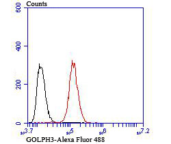
Fig7: Flow cytometric analysis of Hela cells with GOLPH3 antibody at 1/100 dilution (green) compared with an unlabelled control (cells without incubation with primary antibody; red).| Product Name | GOLPH3 |
|---|---|
| Antibody Type | Primary Antibodies |
| Antigen Alias | Coat protein antibody Coat protein GPP34 antibody FLJ90675 antibody Golgi associated protein antibody Golgi peripheral membrane protein 1, 34 kDa antibody Golgi phosphoprotein 3 (coat protein) antibody Golgi phosphoprotein 3 antibody Golgi protein antibody GOLP3_HUMAN antibody Golph3 antibody GOPP1 antibody GPP34 antibody MIDAS antibody Mitochondrial DNA absence factor antibody |
| Product description | GOLPH3 (golgi phosphoprotein 3), also known as GOPP1, GPP34 or MIDAS, is a 298 amino acid protein that localizes to both the cytoplasm and the Golgi stack membrane where it is thought to play a regulatory role in protein trafficking within the Golgi. GOLPH3 is subject to post-translational phosphorylation and is encoded by a gene which maps to human chromosome 5. Chromosome 5 contains 181 million base pairs and comprises nearly 6% of the human genome. Chromosome 5 is associated with Cockayne syndrome through the ERCC8 gene and familial adenomatous polyposis through the adenomatous polyposis coli (APC) tumor suppressor gene. Treacher Collins syndrome is also chromosome 5-associated and is caused by insertions or deletions within the TCOF1 gene. Deletion of the p arm of chromosome 5 leads to Cri du chat syndrome, while deletion of the q arm or of chromosome 5 altogether is common in therapy-related acute myelogenous leukemias and myelodysplastic syndrome. |
| Immunogen | Recombinant protein |
| Clonality | Polyclonal |
|---|---|
| Isotype | IgG |
| Host Species | Rabbit |
| Tested Applications | WBICCIHCFC |
| ICC:1:50-1:200 IHC:1:50-1:200 FC:1:50-1:100 WB:1:500 |
|
| Species Reactivity | HumanMouseRat |
| Concentration | 1mg/ml |
| Alternative Names | Coat protein antibody
Coat protein GPP34 antibody
FLJ90675 antibody
Golgi associated protein antibody
Golgi peripheral membrane protein 1 34 kDa antibody Golgi phosphoprotein 3 (coat protein) antibody Golgi phosphoprotein 3 antibody Golgi protein antibody GOLP3_HUMAN antibody Golph3 antibody GOPP1 antibody GPP34 antibody MIDAS antibody Mitochondrial DNA absence factor antibody |
|---|---|
| Molecular Weight(MW) | 34 kDa |
| Cellular Localization | Cell membrane. Endosome. Cytoplasm. |
Application
Fig1: ICC staining GOLPH3 in Hela cells (green). The nuclear counter stain is DAPI (blue). Cells were fixed in paraformaldehyde, permeabilised with 0.25% Triton X100/PBS.
Application
Fig2: ICC staining GOLPH3 in HepG2 cells (green). The nuclear counter stain is DAPI (blue). Cells were fixed in paraformaldehyde, permeabilised with 0.25% Triton X100/PBS.
Application
Fig3: ICC staining GOLPH3 in SK-Br-3 cells (green). The nuclear counter stain is DAPI (blue). Cells were fixed in paraformaldehyde, permeabilised with 0.25% Triton X100/PBS.
Application
Fig4: Immunohistochemical analysis of paraffin-embedded rat epididymis tissue using anti-GOLPH3 antibody. Counter stained with hematoxylin.
Application
Fig5: Immunohistochemical analysis of paraffin-embedded human stomach cancer tissue using anti-GOLPH3 antibody. Counter stained with hematoxylin.
Application
Fig6: Immunohistochemical analysis of paraffin-embedded mouse stomach tissue using anti-GOLPH3 antibody. Counter stained with hematoxylin.
Application
Fig7: Flow cytometric analysis of Hela cells with GOLPH3 antibody at 1/100 dilution (green) compared with an unlabelled control (cells without incubation with primary antibody; red).| Positive Control | Hela, HepG2, SK-Br-3, rat epididymis tissue, human stomach cancer tissue, mouse stomach tissue. |
|---|---|
| Application Notes | ICC:1:50-1:200 IHC:1:50-1:200 FC:1:50-1:100 WB:1:500 |
| Form | Liquid |
|---|---|
| Storage Instructions | Store at +4℃ after thawing. Aliquot store at -20℃ or -80℃. Avoid repeated freeze / thaw cycles. |
| Storage Buffer | 1*TBS (pH7.4), 0.5%BSA, 50%Glycerol. Preservative: 0.05% Sodium Azide. |


