Application
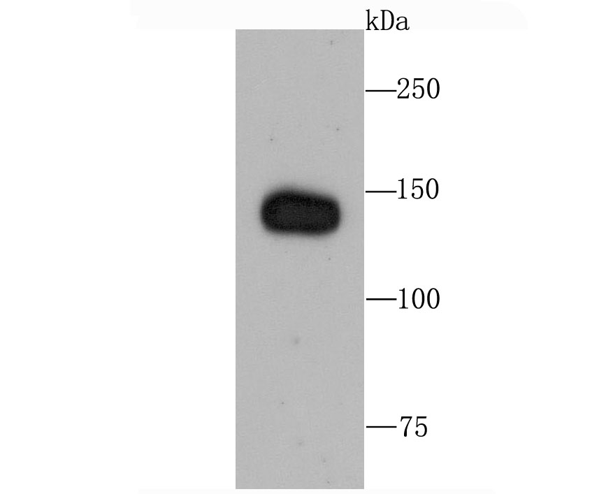
Fig1: Western blot analysis of Factor H on human lung tissue lysate using anti-Factor H antibody at 1/1,000 dilution.Application
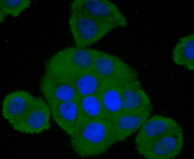
Fig2: ICC staining Factor H in Hela cells (green). The nuclear counter stain is DAPI (blue). Cells were fixed in paraformaldehyde, permeabilised with 0.25% Triton X100/PBS.Application
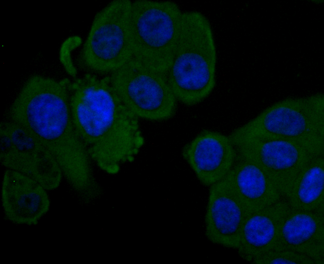
Fig3: ICC staining Factor H in HepG2 cells (green). The nuclear counter stain is DAPI (blue). Cells were fixed in paraformaldehyde, permeabilised with 0.25% Triton X100/PBS.| Product Name | Factor H [JA52-13] |
|---|---|
| Antibody Type | Primary Antibodies |
| Antigen Alias | adrenomedullin binding protein antibody age related maculopathy susceptibility 1 antibody AHUS 1 antibody AHUS1 antibody AMBP 1 antibody AMBP1 antibody ARMD 4 antibody ARMD4 antibody ARMS 1 antibody ARMS1 antibody beta 1 H globulin antibody beta 1H antibody beta1H antibody CFAH_HUMAN antibody CFH antibody CFHL 3 antibody CFHL3 antibody Complement factor H antibody complement factor H, isoform b antibody Factor H antibody factor H like 1 antibody FH antibody FHL 1 antibody FHL1 antibody H factor 1 (complement) antibody H factor 1 antibody H factor 2 (complement) antibody HF 1 antibody HF 2 antibody HF antibody HF1 antibody HF2 antibody HUS antibody MGC88246 antibody |
| Product description | The Factor H gene family is a multidomain, multifunctional protein family whose individual members are defined by conserved structural elements, which display diverse yet often overlapping functions. These proteins share a common structural motif, the short consensus repeat (SCR), which is structurally conserved among related genes and between phylogenetically divergent species. The human complement Factor H (FH, CFH, HUS, b-1H) gene encodes a 1213 amino acid serum glycoprotein which is arranged into 20 SCRs, each approximately 60 amino acids long, and an 18-residue leader sequence. Factor H controls the function of the alternative complement pathway and acts as a cofactor with Factor I (C3b inactivator). In addition, Factor H has functional activity outside of the complement system, where it can bind to the cellular integrin receptor (CD11b/CD18), interact with cell surface glycosaminoglycans and associate with the surface of certain pathogenic microorganisms. Deficiencies in Factor H is a common characteristic of acute renal disease. |
| Immunogen | recombinant protein |
| Clonality | Monoclonal |
|---|---|
| Isotype | IgG |
| Host Species | Recombinant rabbit |
| Tested Applications | WBICC |
| WB:1:500-1:2,000 ICC:1:50-1:200 |
|
| Species Reactivity | Human |
| Concentration | 1mg/ml |
| Alternative Names | adrenomedullin binding protein antibody
age related maculopathy susceptibility 1 antibody
AHUS 1 antibody
AHUS1 antibody
AMBP 1 antibody
AMBP1 antibody
ARMD 4 antibody
ARMD4 antibody
ARMS 1 antibody
ARMS1 antibody
beta 1 H globulin antibody
beta 1H antibody
beta1H antibody
CFAH_HUMAN antibody
CFH antibody
CFHL 3 antibody
CFHL3 antibody
Complement factor H antibody
complement factor H isoform b antibody Factor H antibody factor H like 1 antibody FH antibody FHL 1 antibody FHL1 antibody H factor 1 (complement) antibody H factor 1 antibody H factor 2 (complement) antibody HF 1 antibody HF 2 antibody HF antibody HF1 antibody HF2 antibody HUS antibody MGC88246 antibody |
|---|---|
| Molecular Weight(MW) | 140 kDa |
| Cellular Localization | Secreted. |
| SwissProt ID | P08603 |
|---|
Application
Fig1: Western blot analysis of Factor H on human lung tissue lysate using anti-Factor H antibody at 1/1,000 dilution.
Application
Fig2: ICC staining Factor H in Hela cells (green). The nuclear counter stain is DAPI (blue). Cells were fixed in paraformaldehyde, permeabilised with 0.25% Triton X100/PBS.
Application
Fig3: ICC staining Factor H in HepG2 cells (green). The nuclear counter stain is DAPI (blue). Cells were fixed in paraformaldehyde, permeabilised with 0.25% Triton X100/PBS.| Positive Control | Human lung tissue lysate, Hela, HepG2. |
|---|---|
| Application Notes | WB:1:500-1:2,000 ICC:1:50-1:200 |
| Form | Liquid |
|---|---|
| Storage Instructions | Store at +4℃ after thawing. Aliquot store at -20℃ or -80℃. Avoid repeated freeze / thaw cycles. |
| Storage Buffer | 1*TBS (pH7.4), 1%BSA, 40%Glycerol. Preservative: 0.05% Sodium Azide. |


