Application
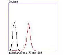
Fig6: Flow cytometric analysis of MCF-7 cells with mSin3A antibody at 1/100 dilution (red) compared with an unlabelled control (cells without incubation with primary antibody; black).Application
Fig6: Flow cytometric analysis of MCF-7 cells with mSin3A antibody at 1/100 dilution (red) compared with an unlabelled control (cells without incubation with primary antibody; black).Application
Fig6: Flow cytometric analysis of MCF-7 cells with mSin3A antibody at 1/100 dilution (red) compared with an unlabelled control (cells without incubation with primary antibody; black).Application
Fig6: Flow cytometric analysis of MCF-7 cells with mSin3A antibody at 1/100 dilution (red) compared with an unlabelled control (cells without incubation with primary antibody; black).Application
Fig6: Flow cytometric analysis of MCF-7 cells with mSin3A antibody at 1/100 dilution (red) compared with an unlabelled control (cells without incubation with primary antibody; black).Application
Fig6: Flow cytometric analysis of MCF-7 cells with mSin3A antibody at 1/100 dilution (red) compared with an unlabelled control (cells without incubation with primary antibody; black).| Product Name | mSin3A [JA94-31] |
|---|---|
| Antibody Type | Primary Antibodies |
| Antigen Alias | AW553200 antibody DKFZP434K2235 antibody FLJ90319 antibody Histone deacetylase complex subunit Sin 3a antibody Histone deacetylase complex subunit Sin3a antibody KIAA0700 antibody Kiaa4126 antibody mKIAA4126 antibody Paired amphipathic helix protein Sin 3a antibody Paired amphipathic helix protein Sin3a antibody Sin 3a antibody SIN3 homolog A antibody SIN3 homolog A transcription regulator (yeast) antibody SIN3 homolog A transcription regulator antibody SIN3 transcription regulator homolog A antibody Sin3a antibody SIN3A protein antibody SIN3A_HUMAN antibody Transcriptional co repressor Sin 3A antibody Transcriptional co repressor Sin3A antibody Transcriptional corepressor Sin3a antibody Transcriptional regulator SIN3A antibody |
| Product description | It is now well established that Myc regulation of cell proliferation and differentiation involves a family of related transcription factors. One such factor, Max, is an obligate heterodimeric partner for Myc and can also form heterodimers with at least four related proteins designated Mad 1, Mxi1 (alternatively designated Mad 2), Mad 3 and Mad 4. Like Mad 1 and Mxi1, association of Mad 3 and Mad 4 with Max results in transcriptional repression. Both Myc and the Mad proteins have short half-lives and their synthesis is tightly regulated, while Max expression is constitutive and relatively stable. Two related mammalian cDNAs have been identified and shown to encode Mad-binding proteins. Both possess sequence homology with the yeast transcription repressor Sin3 including four conserved paired amphipathic helix (PAH) domains. mSin3A and mSin3B specifically interact with the Mad proteins via their second paired amphipathic helix domain (PAH2). It has been suggested that Mad-Max heterodimers repress transcription by tethering mSin3 to DNA as corepressors. |
| Immunogen | recombinant protein |
| Clonality | Monoclonal |
|---|---|
| Isotype | IgG |
| Host Species | Recombinant rabbit |
| Tested Applications | WBICC/IFIHCFC |
| WB:1:500-1:2,000 ICC:1:50-1:200 IHC:1:50 FC:1:50-1:100 |
|
| Species Reactivity | HumanMouseRat |
| Concentration | 1mg/ml |
| Alternative Names | AW553200 antibody DKFZP434K2235 antibody FLJ90319 antibody Histone deacetylase complex subunit Sin 3a antibody Histone deacetylase complex subunit Sin3a antibody KIAA0700 antibody Kiaa4126 antibody mKIAA4126 antibody Paired amphipathic helix protein Sin 3a antibody Paired amphipathic helix protein Sin3a antibody Sin 3a antibody SIN3 homolog A antibody SIN3 homolog A transcription regulator (yeast) antibody SIN3 homolog A transcription regulator antibody SIN3 transcription regulator homolog A antibody Sin3a antibody SIN3A protein antibody SIN3A_HUMAN antibody Transcriptional co repressor Sin 3A antibody Transcriptional co repressor Sin3A antibody Transcriptional corepressor Sin3a antibody Transcriptional regulator SIN3A antibody |
|---|---|
| Molecular Weight(MW) | 145 kDa |
| Cellular Localization | Nucleus. |
Application
Fig6: Flow cytometric analysis of MCF-7 cells with mSin3A antibody at 1/100 dilution (red) compared with an unlabelled control (cells without incubation with primary antibody; black).
Application
Fig6: Flow cytometric analysis of MCF-7 cells with mSin3A antibody at 1/100 dilution (red) compared with an unlabelled control (cells without incubation with primary antibody; black).
Application
Fig6: Flow cytometric analysis of MCF-7 cells with mSin3A antibody at 1/100 dilution (red) compared with an unlabelled control (cells without incubation with primary antibody; black).
Application
Fig6: Flow cytometric analysis of MCF-7 cells with mSin3A antibody at 1/100 dilution (red) compared with an unlabelled control (cells without incubation with primary antibody; black).
Application
Fig6: Flow cytometric analysis of MCF-7 cells with mSin3A antibody at 1/100 dilution (red) compared with an unlabelled control (cells without incubation with primary antibody; black).
Application
Fig6: Flow cytometric analysis of MCF-7 cells with mSin3A antibody at 1/100 dilution (red) compared with an unlabelled control (cells without incubation with primary antibody; black).| Positive Control | 293T, Hela, MCF-7, NIH-3T3, human kidney tissue. |
|---|---|
| Application Notes | WB:1:500-1:2,000 ICC:1:50-1:200 IHC:1:50 FC:1:50-1:100 |
| Form | Liquid |
|---|---|
| Storage Instructions | Store at +4℃ after thawing. Aliquot store at -20℃ or -80℃. Avoid repeated freeze / thaw cycles. |
| Storage Buffer | 1*TBS (pH7.4), 1%BSA, 40%Glycerol. Preservative: 0.05% Sodium Azide. |


