Application
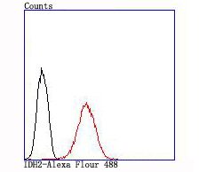
Fig7: Flow cytometric analysis of MCF-7 cells with IDH2 antibody at 1/100 dilution (red) compared with an unlabelled control (cells without incubation with primary antibody; black).Application
Fig7: Flow cytometric analysis of MCF-7 cells with IDH2 antibody at 1/100 dilution (red) compared with an unlabelled control (cells without incubation with primary antibody; black).Application
Fig7: Flow cytometric analysis of MCF-7 cells with IDH2 antibody at 1/100 dilution (red) compared with an unlabelled control (cells without incubation with primary antibody; black).Application
Fig7: Flow cytometric analysis of MCF-7 cells with IDH2 antibody at 1/100 dilution (red) compared with an unlabelled control (cells without incubation with primary antibody; black).Application
Fig7: Flow cytometric analysis of MCF-7 cells with IDH2 antibody at 1/100 dilution (red) compared with an unlabelled control (cells without incubation with primary antibody; black).Application
Fig7: Flow cytometric analysis of MCF-7 cells with IDH2 antibody at 1/100 dilution (red) compared with an unlabelled control (cells without incubation with primary antibody; black).Application
Fig7: Flow cytometric analysis of MCF-7 cells with IDH2 antibody at 1/100 dilution (red) compared with an unlabelled control (cells without incubation with primary antibody; black).| Product Name | IDH2 [JA55-31] |
|---|---|
| Antibody Type | Primary Antibodies |
| Antigen Alias | D2HGA2 antibody ICD-M antibody IDH antibody IDH2 antibody IDHM antibody IDHP_HUMAN antibody IDP antibody IDPM antibody Isocitrate dehydrogenase [NADP], mitochondrial antibody Isocitrate dehydrogenase 2 (NADP+), mitochondrial antibody mNADP-IDH antibody NADP(+)-specific ICDH antibody Oxalosuccinate decarboxylase antibody |
| Product description | IDH2 (isocitrate dehydrogenase 2 (NADP+), mitochondrial), also designated NADP+-specific ICDH; isocitrate dehydrogenase, mitochondrial; and oxalosuccinate decarboxylase, is a 452 amino acid enzyme encoded by the human gene IDH2. IDH2 belongs to the isocitrate and isopropylmalate dehydrogenases family and contains two nucleotide binding regions. IDH2 is involved in the reduction of NADP+ to NADPH and maintains the supply of glutathione (GSH) in mitochondria. It is believed to play a role in intermediary metabolism and energy production. IDH2 also tightly associates with the pyruvate dehydrogenase complex. IDH2 is found in the mitochondrion as a homodimer and can bind one magnesium or manganese ion per subunit. |
| Immunogen | recombinant protein |
| Clonality | Monoclonal |
|---|---|
| Isotype | IgG |
| Host Species | Recombinant rabbit |
| Tested Applications | WBIHCFC |
| WB:1:500-1:2,000 IHC:1:50-1:200 FC:1:50-1:100 |
|
| Species Reactivity | HumanMouseRat |
| Concentration | 1mg/ml |
| Alternative Names | D2HGA2 antibody
ICD-M antibody
IDH antibody
IDH2 antibody
IDHM antibody
IDHP_HUMAN antibody
IDP antibody
IDPM antibody
Isocitrate dehydrogenase [NADP] mitochondrial antibody Isocitrate dehydrogenase 2 (NADP+) mitochondrial antibody mNADP-IDH antibody NADP(+)-specific ICDH antibody Oxalosuccinate decarboxylase antibody |
|---|---|
| Molecular Weight(MW) | 45 kDa |
| Cellular Localization | Mitochondrion. |
Application
Fig7: Flow cytometric analysis of MCF-7 cells with IDH2 antibody at 1/100 dilution (red) compared with an unlabelled control (cells without incubation with primary antibody; black).
Application
Fig7: Flow cytometric analysis of MCF-7 cells with IDH2 antibody at 1/100 dilution (red) compared with an unlabelled control (cells without incubation with primary antibody; black).
Application
Fig7: Flow cytometric analysis of MCF-7 cells with IDH2 antibody at 1/100 dilution (red) compared with an unlabelled control (cells without incubation with primary antibody; black).
Application
Fig7: Flow cytometric analysis of MCF-7 cells with IDH2 antibody at 1/100 dilution (red) compared with an unlabelled control (cells without incubation with primary antibody; black).
Application
Fig7: Flow cytometric analysis of MCF-7 cells with IDH2 antibody at 1/100 dilution (red) compared with an unlabelled control (cells without incubation with primary antibody; black).
Application
Fig7: Flow cytometric analysis of MCF-7 cells with IDH2 antibody at 1/100 dilution (red) compared with an unlabelled control (cells without incubation with primary antibody; black).
Application
Fig7: Flow cytometric analysis of MCF-7 cells with IDH2 antibody at 1/100 dilution (red) compared with an unlabelled control (cells without incubation with primary antibody; black).| Positive Control | 293T, mouse heart tissue lysate, rat heart tissue lysate, human liver tissue, human prostate tissue, MCF-7. |
|---|---|
| Application Notes | WB:1:500-1:2,000 IHC:1:50-1:200 FC:1:50-1:100 |
| Form | Liquid |
|---|---|
| Storage Instructions | Store at +4℃ after thawing. Aliquot store at -20℃ or -80℃. Avoid repeated freeze / thaw cycles. |
| Storage Buffer | 1*TBS (pH7.4), 1%BSA, 40%Glycerol. Preservative: 0.05% Sodium Azide. |


