Application
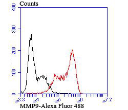
Fig7: Flow cytometric analysis of HL-60 cells with MMP9 antibody at 1/100 dilution (red) compared with an unlabelled control (cells without incubation with primary antibody; black). Goat anti rabbit IgG (FITC) was used as the secondary antibodyApplication
Fig7: Flow cytometric analysis of HL-60 cells with MMP9 antibody at 1/100 dilution (red) compared with an unlabelled control (cells without incubation with primary antibody; black). Goat anti rabbit IgG (FITC) was used as the secondary antibodyApplication
Fig7: Flow cytometric analysis of HL-60 cells with MMP9 antibody at 1/100 dilution (red) compared with an unlabelled control (cells without incubation with primary antibody; black). Goat anti rabbit IgG (FITC) was used as the secondary antibodyApplication
Fig7: Flow cytometric analysis of HL-60 cells with MMP9 antibody at 1/100 dilution (red) compared with an unlabelled control (cells without incubation with primary antibody; black). Goat anti rabbit IgG (FITC) was used as the secondary antibodyApplication
Fig7: Flow cytometric analysis of HL-60 cells with MMP9 antibody at 1/100 dilution (red) compared with an unlabelled control (cells without incubation with primary antibody; black). Goat anti rabbit IgG (FITC) was used as the secondary antibodyApplication
Fig7: Flow cytometric analysis of HL-60 cells with MMP9 antibody at 1/100 dilution (red) compared with an unlabelled control (cells without incubation with primary antibody; black). Goat anti rabbit IgG (FITC) was used as the secondary antibodyApplication
Fig7: Flow cytometric analysis of HL-60 cells with MMP9 antibody at 1/100 dilution (red) compared with an unlabelled control (cells without incubation with primary antibody; black). Goat anti rabbit IgG (FITC) was used as the secondary antibody| Product Name | MMP9 |
|---|---|
| Antibody Type | Primary Antibodies |
| Antigen Alias | 82 kDa matrix metalloproteinase-9 antibody 92 kDa gelatinase antibody 92 kDa type IV collagenase antibody CLG 4B antibody CLG4B antibody Collagenase Type 4 beta antibody Collagenase type IV 92 KD antibody EC 3.4.24.35 antibody Gelatinase 92 KD antibody Gelatinase B antibody Gelatinase beta antibody GelatinaseB antibody GELB antibody Macrophage gelatinase antibody MANDP2 antibody Matrix metallopeptidase 9 (gelatinase B, 92kDa gelatinase, 92kDa type IV collagenase) antibody Matrix Metalloproteinase 9 antibody MMP 9 antibody MMP-9 antibody MMP9 antibody MMP9_HUMAN antibody Type V collagenase antibody |
| Product description | The matrix metalloproteinases (MMP) are a family of peptidase enzymes responsible for the degradation of extracellular matrix components, including collagen, gelatin, fibronectin, laminin and proteoglycan. Transcription of MMP genes is differentially activated by phorbol ester, lipopolysaccharide (LPS) or staphylococcal enterotoxin B (SEB). MMP catalysis requires both calcium and zinc. MMP-9 (also designated 92 kDa type IV collagenase or gelatinase B) has been shown to degrade bone collagens in concert with MMP-1 (also designated interstitial collagenase, fibroblast collagenase or collagenase-1), and cysteine proteases and may play a role in bone osteoclastic resorption. MMP-1 is downregulated by p53, and abnormality of p53 expression may contribute to joint degradation in rheumatoid arthritis by regulating MMP-1 expression. |
| Immunogen | Peptide |
| Clonality | Polyclonal |
|---|---|
| Isotype | IgG |
| Host Species | Rabbit |
| Tested Applications | WBICCIHCFC |
| ICC:1:50-1:200 IHC:1:50-1:200 FC:1:50-1:100 WB:1:500 |
|
| Species Reactivity | HumanMouse |
| Concentration | 1mg/ml |
| Alternative Names | 82 kDa matrix metalloproteinase-9 antibody
92 kDa gelatinase antibody
92 kDa type IV collagenase antibody
CLG 4B antibody
CLG4B antibody
Collagenase Type 4 beta antibody
Collagenase type IV 92 KD antibody
EC 3.4.24.35 antibody
Gelatinase 92 KD antibody
Gelatinase B antibody
Gelatinase beta antibody
GelatinaseB antibody
GELB antibody
Macrophage gelatinase antibody
MANDP2 antibody
Matrix metallopeptidase 9 (gelatinase B 92kDa gelatinase 92kDa type IV collagenase) antibody Matrix Metalloproteinase 9 antibody MMP 9 antibody MMP-9 antibody MMP9 antibody MMP9_HUMAN antibody Type V collagenase antibody |
|---|---|
| Molecular Weight(MW) | 100 kDa |
| Cellular Localization | Extracellular matrix. Secreted. |
Application
Fig7: Flow cytometric analysis of HL-60 cells with MMP9 antibody at 1/100 dilution (red) compared with an unlabelled control (cells without incubation with primary antibody; black). Goat anti rabbit IgG (FITC) was used as the secondary antibody
Application
Fig7: Flow cytometric analysis of HL-60 cells with MMP9 antibody at 1/100 dilution (red) compared with an unlabelled control (cells without incubation with primary antibody; black). Goat anti rabbit IgG (FITC) was used as the secondary antibody
Application
Fig7: Flow cytometric analysis of HL-60 cells with MMP9 antibody at 1/100 dilution (red) compared with an unlabelled control (cells without incubation with primary antibody; black). Goat anti rabbit IgG (FITC) was used as the secondary antibody
Application
Fig7: Flow cytometric analysis of HL-60 cells with MMP9 antibody at 1/100 dilution (red) compared with an unlabelled control (cells without incubation with primary antibody; black). Goat anti rabbit IgG (FITC) was used as the secondary antibody
Application
Fig7: Flow cytometric analysis of HL-60 cells with MMP9 antibody at 1/100 dilution (red) compared with an unlabelled control (cells without incubation with primary antibody; black). Goat anti rabbit IgG (FITC) was used as the secondary antibody
Application
Fig7: Flow cytometric analysis of HL-60 cells with MMP9 antibody at 1/100 dilution (red) compared with an unlabelled control (cells without incubation with primary antibody; black). Goat anti rabbit IgG (FITC) was used as the secondary antibody
Application
Fig7: Flow cytometric analysis of HL-60 cells with MMP9 antibody at 1/100 dilution (red) compared with an unlabelled control (cells without incubation with primary antibody; black). Goat anti rabbit IgG (FITC) was used as the secondary antibody| Positive Control | Hela, MCF-7, PANC-1, human tonsil tissue, human spleen tissue, mouse spleen tissue, HL-60. |
|---|---|
| Application Notes | ICC:1:50-1:200 IHC:1:50-1:200 FC:1:50-1:100 WB:1:500 |
| Form | Liquid |
|---|---|
| Storage Instructions | Store at +4℃ after thawing. Aliquot store at -20℃ or -80℃. Avoid repeated freeze / thaw cycles. |
| Storage Buffer | 1*TBS (pH7.4), 0.5%BSA, 50%Glycerol. Preservative: 0.05% Sodium Azide. |


