Application
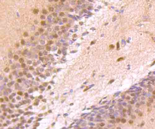
Fig1: Immunohistochemical analysis of paraffin-embedded rat brain tissue using anti-Zic1 antibody. Counter stained with hematoxylin.Application
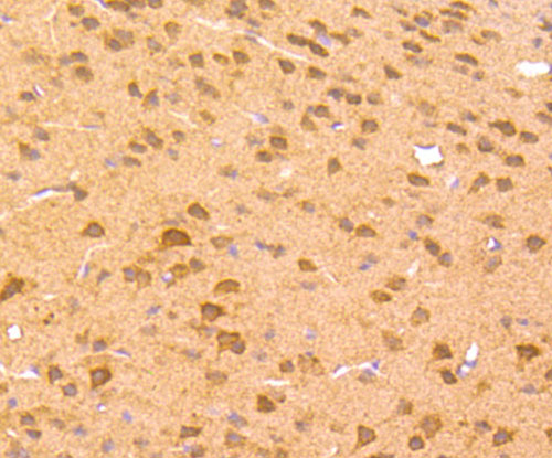
Fig2: Immunohistochemical analysis of paraffin-embedded mouse brain tissue using anti-Zic1 antibody. Counter stained with hematoxylin.Application
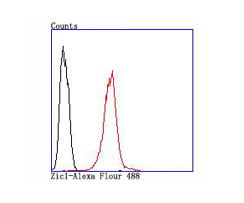
Fig3: Flow cytometric analysis of SH-SY5Y cells with Zic1 antibody at 1/100 dilution (red) compared with an unlabelled control (cells without incubation with primary antibody; black).| Product Name | Zic1 [JA31-60] |
|---|---|
| Antibody Type | Primary Antibodies |
| Antigen Alias | Odd paired homolog Drosophila antibody Zic 1 antibody ZIC antibody Zic family member 1 (odd-paired Drosophila homolog) antibody Zic family member 1 antibody Zic protein member 1 antibody zic1 antibody ZIC1_HUMAN antibody Zinc finger protein 201 antibody Zinc finger protein of the cerebellum 1 antibody Zinc finger protein ZIC 1 antibody Zinc finger protein ZIC1 antibody ZNF 201 antibody ZNF201 antibody |
| Product description | Zic1 (zinc finger protein of the cerebellum 1) is a C2H2 zinc finger transcription factor that controls the expansion of neuronal precursors by inhibiting the progression of neuronal differentiation. Zic1 determines the cerebellar folial pattern by influencing proliferation in the external germinal layer (EGL). Zic1 can bind and transactivate the apolipoprotein E gene.This gene is closely linked to the gene encoding zinc finger protein of the cerebellum 4, a related family member on chromosome 3. |
| Immunogen | Recombinant protein |
| Clonality | Monoclonal |
|---|---|
| Isotype | IgG |
| Host Species | Recombinant rabbit |
| Tested Applications | WBIHCFC |
| WB:1:500~1:2000 IHC:1:50~1:200 FC:1:200~1:500 Notes:Optimal dilutions/concentrations should be determined by the researcher. |
|
| Species Reactivity | HumanMouseRat |
| Concentration | 1mg/ml |
| Alternative Names | Odd paired homolog Drosophila antibody Zic 1 antibody ZIC antibody Zic family member 1 (odd-paired Drosophila homolog) antibody Zic family member 1 antibody Zic protein member 1 antibody zic1 antibody ZIC1_HUMAN antibody Zinc finger protein 201 antibody Zinc finger protein of the cerebellum 1 antibody Zinc finger protein ZIC 1 antibody Zinc finger protein ZIC1 antibody ZNF 201 antibody ZNF201 antibody |
|---|---|
| Molecular Weight(MW) | 48 kDa |
| Cellular Localization | Nucleus. Cytoplasm. |
Application
Fig1: Immunohistochemical analysis of paraffin-embedded rat brain tissue using anti-Zic1 antibody. Counter stained with hematoxylin.
Application
Fig2: Immunohistochemical analysis of paraffin-embedded mouse brain tissue using anti-Zic1 antibody. Counter stained with hematoxylin.
Application
Fig3: Flow cytometric analysis of SH-SY5Y cells with Zic1 antibody at 1/100 dilution (red) compared with an unlabelled control (cells without incubation with primary antibody; black).| Positive Control | Rat brain tissue, mouse brain tissue, SH-SY5Y. |
|---|---|
| Application Notes | WB:1:500~1:2000 IHC:1:50~1:200 FC:1:200~1:500 Notes:Optimal dilutions/concentrations should be determined by the researcher. |
| Form | Liquid |
|---|---|
| Storage Instructions | Store at +4℃ after thawing. Aliquot store at -20℃ or -80℃. Avoid repeated freeze / thaw cycles. |
| Storage Buffer | 1*TBS (pH7.4), 1%BSA, 40%Glycerol. Preservative: 0.05% Sodium Azide. |


