Application
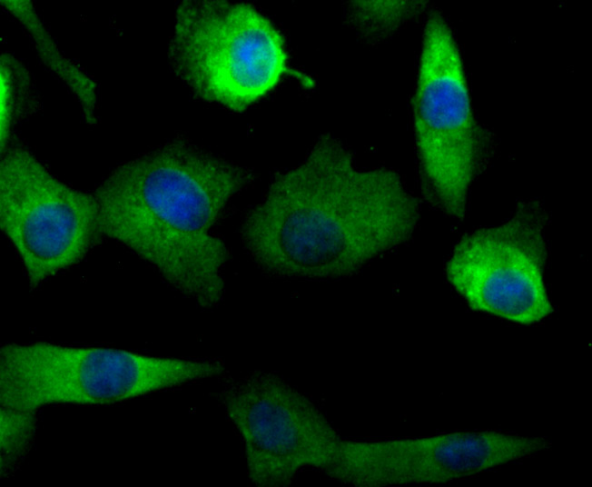
Fig1: ICC staining NaV1.7 in A549 cells (green). The nuclear counter stain is DAPI (blue). Cells were fixed in paraformaldehyde, permeabilised with 0.25% Triton X100/PBS.Application
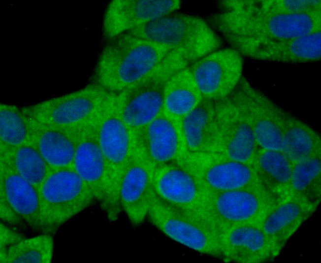
Fig2: ICC staining NaV1.7 in Hela cells (green). The nuclear counter stain is DAPI (blue). Cells were fixed in paraformaldehyde, permeabilised with 0.25% Triton X100/PBS.Application
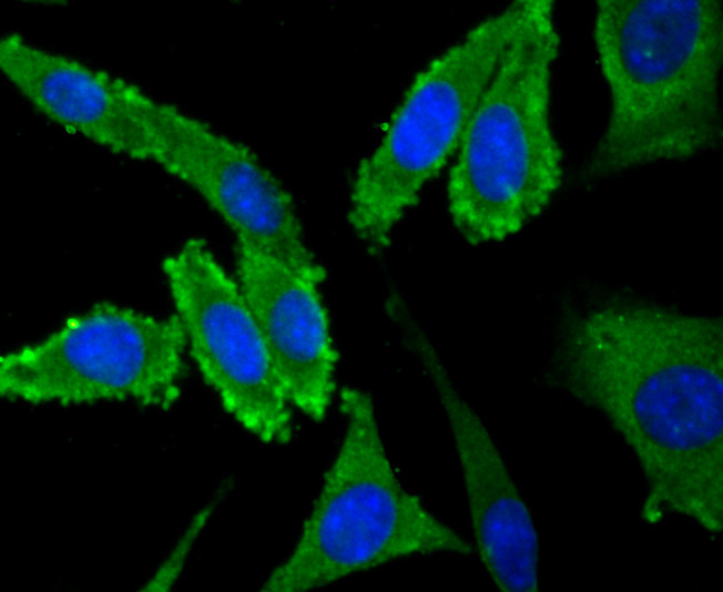
Fig3: ICC staining NaV1.7 in SH-SY5Y cells (green). The nuclear counter stain is DAPI (blue). Cells were fixed in paraformaldehyde, permeabilised with 0.25% Triton X100/PBS.Application
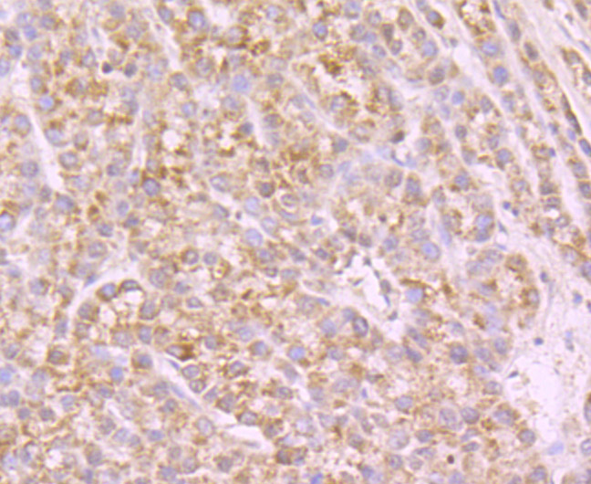
Fig4: Immunohistochemical analysis of paraffin-embedded human liver cancer tissue using anti-NaV1.7 beta antibody. Counter stained with hematoxylin.Application
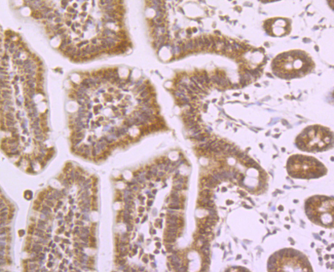
Fig5: Immunohistochemical analysis of paraffin-embedded mouse colon tissue using anti-NaV1.7 beta antibody. Counter stained with hematoxylin.Application
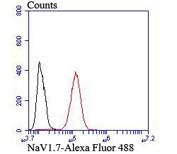
Fig6: Flow cytometric analysis of SH-SY5Y cells with NaV1.7 antibody at 1/100 dilution (red) compared with an unlabelled control (cells without incubation with primary antibody; black).| Product Name | NaV1.7 |
|---|---|
| Antibody Type | Primary Antibodies |
| Antigen Alias | ETHA antibody GEFSP7 antibody hNE Na antibody hNE-Na antibody hNENa antibody NE NA antibody NENA antibody Neuroendocrine sodium channel antibody Peripheral sodium channel 1 antibody PN1 antibody Scn9a antibody SCN9A_HUMAN antibody Sodium channel protein type 9 subunit alpha antibody Sodium channel protein type IX subunit alpha antibody Sodium channel voltage gated type IX alpha antibody Sodium channel voltage gated type IX alpha polypeptide antibody Sodium channel voltage gated type IX alpha subunit antibody Voltage gated sodium channel alpha subunit Nav1.7 antibody Voltage gated sodium channel subunit alpha Nav1 antibody Voltage-gated sodium channel subunit alpha Nav1.7 antibody |
| Product description | Mediates the voltage-dependent sodium ion permeability of excitable membranes. Assuming opened or closed conformations in response to the voltage difference across the membrane, the protein forms a sodium-selective channel through which Na+ ions may pass in accordance with their electrochemical gradient. It is a tetrodotoxin-sensitive Na+ channel isoform. Plays a role in pain mechanisms, especially in the development of inflammatory pain. |
| Immunogen | Peptide |
| Clonality | Polyclonal |
|---|---|
| Isotype | IgG |
| Host Species | Rabbit |
| Tested Applications | ICCIHCFC |
| ICC:1:50-1:200 ICC:1:50-1:200 FC:1:50-1:100 |
|
| Species Reactivity | Human |
| Concentration | 1mg/ml |
| Alternative Names | ETHA antibody GEFSP7 antibody hNE Na antibody hNE-Na antibody hNENa antibody NE NA antibody NENA antibody Neuroendocrine sodium channel antibody Peripheral sodium channel 1 antibody PN1 antibody Scn9a antibody SCN9A_HUMAN antibody Sodium channel protein type 9 subunit alpha antibody Sodium channel protein type IX subunit alpha antibody Sodium channel voltage gated type IX alpha antibody Sodium channel voltage gated type IX alpha polypeptide antibody Sodium channel voltage gated type IX alpha subunit antibody Voltage gated sodium channel alpha subunit Nav1.7 antibody Voltage gated sodium channel subunit alpha Nav1 antibody Voltage-gated sodium channel subunit alpha Nav1.7 antibody |
|---|---|
| Molecular Weight(MW) | 226 kDa |
| Cellular Localization | Cell membrane. Cell projection. |
| SwissProt ID | Q15858 |
|---|
Application
Fig1: ICC staining NaV1.7 in A549 cells (green). The nuclear counter stain is DAPI (blue). Cells were fixed in paraformaldehyde, permeabilised with 0.25% Triton X100/PBS.
Application
Fig2: ICC staining NaV1.7 in Hela cells (green). The nuclear counter stain is DAPI (blue). Cells were fixed in paraformaldehyde, permeabilised with 0.25% Triton X100/PBS.
Application
Fig3: ICC staining NaV1.7 in SH-SY5Y cells (green). The nuclear counter stain is DAPI (blue). Cells were fixed in paraformaldehyde, permeabilised with 0.25% Triton X100/PBS.
Application
Fig4: Immunohistochemical analysis of paraffin-embedded human liver cancer tissue using anti-NaV1.7 beta antibody. Counter stained with hematoxylin.
Application
Fig5: Immunohistochemical analysis of paraffin-embedded mouse colon tissue using anti-NaV1.7 beta antibody. Counter stained with hematoxylin.
Application
Fig6: Flow cytometric analysis of SH-SY5Y cells with NaV1.7 antibody at 1/100 dilution (red) compared with an unlabelled control (cells without incubation with primary antibody; black).| Positive Control | A549, Hela, SH-SY5Y, human liver cancer tissue, mouse colon tissue. |
|---|---|
| Application Notes | ICC:1:50-1:200 ICC:1:50-1:200 FC:1:50-1:100 |
| Form | Liquid |
|---|---|
| Storage Instructions | Store at +4℃ after thawing. Aliquot store at -20℃ or -80℃. Avoid repeated freeze / thaw cycles. |
| Storage Buffer | 1*TBS (pH7.4), 0.5%BSA, 50%Glycerol. Preservative: 0.05% Sodium Azide. |


