Application
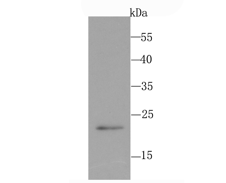
Fig1: Western blot analysis of PMP22 on PC-12 cell using anti-PMP22 antibody at 1/500 dilution.Application
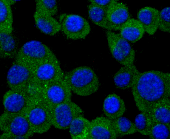
Fig2: ICC staining PMP22 in N2A cells (green). The nuclear counter stain is DAPI (blue). Cells were fixed in paraformaldehyde, permeabilised with 0.25% Triton X100/PBS.Application
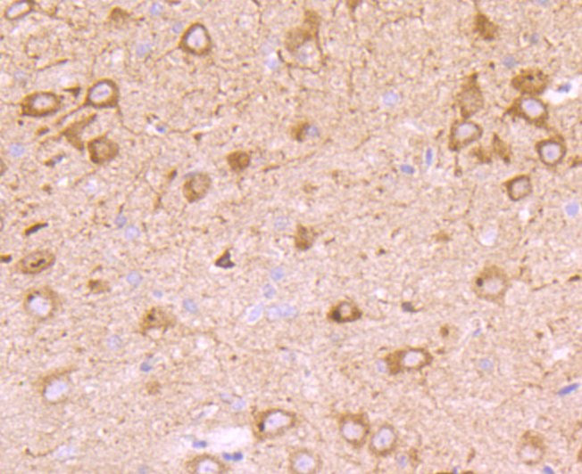
Fig3: Immunohistochemical analysis of paraffin-embedded rat brain tissue using anti-PMP22 antibody. Counter stained with hematoxylin.Application
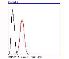
Fig4: Flow cytometric analysis of SH-SY5Y cells with PMP22 antibody at 1/100 dilution (red) compared with an unlabelled control (cells without incubation with primary antibody; black).| Product Name | PMP22 [JM52-30] |
|---|---|
| Antibody Type | Primary Antibodies |
| Antigen Alias | CMT1A antibody CMT1E antibody DSS antibody GAS-3 antibody GAS3 antibody Growth Arrest Specific 3 antibody Growth arrest-specific protein 3 antibody HMSNIA antibody HNPP antibody MGC20769 antibody Peripheral myelin protein 22 antibody PMP-22 antibody PMP22 antibody PMP22_HUMAN antibody Sp110 antibody Trembler antibody |
| Product description | PLP (myelin proteolipid protein or lipophilin) is a major constituent of myelin. The two isoforms of the myelin proteolipid protein, PLP and DM20, are very hydrophobic integral membrane proteins that account for about half of the protein content of adult CNS myelin. A mutation in the gene which encodes PLP is linked to Pelizaeus-Merzbacher disease (PMD), a chronic infantile type of diffuse cerebral sclerosis. The gene which encodes PLP maps to human chromosome Xq13-q22. The glycoprotein zero (also designated P-zero or myelin peripheral protein) is the major structural protein of peripheral myelin, accounting for more than 50% of the protein present in the sheath of peri-pheral nerves. Zero is an integral membrane glycoprotein whose expression is restricted to Schwann cells. The gene which encodes zero maps to human chromosome 1q22. PMP22 (peripheral myelin protein 22) is a growth-regulated membrane protein which is expressed by Schwann cells and is localized mainly in compact peripheral nervous system myelin. The gene which encodes PMP22 maps to human chromosome 17p11.2. |
| Immunogen | recombinant protein |
| Clonality | Monoclonal |
|---|---|
| Isotype | IgG |
| Host Species | Recombinant rabbit |
| Tested Applications | WBICCIHCFC |
| WB:1:500 ICC:1:50-1:200 IHC:1:50-1:200 FC:1:50-1:100 |
|
| Species Reactivity | HumanRat |
| Concentration | 1mg/ml |
| Alternative Names | CMT1A antibody CMT1E antibody DSS antibody GAS-3 antibody GAS3 antibody Growth Arrest Specific 3 antibody Growth arrest-specific protein 3 antibody HMSNIA antibody HNPP antibody MGC20769 antibody Peripheral myelin protein 22 antibody PMP-22 antibody PMP22 antibody PMP22_HUMAN antibody Sp110 antibody Trembler antibody |
|---|---|
| Molecular Weight(MW) | 22 kDa |
| Cellular Localization | Membrane. |
Application
Fig1: Western blot analysis of PMP22 on PC-12 cell using anti-PMP22 antibody at 1/500 dilution.
Application
Fig2: ICC staining PMP22 in N2A cells (green). The nuclear counter stain is DAPI (blue). Cells were fixed in paraformaldehyde, permeabilised with 0.25% Triton X100/PBS.
Application
Fig3: Immunohistochemical analysis of paraffin-embedded rat brain tissue using anti-PMP22 antibody. Counter stained with hematoxylin.
Application
Fig4: Flow cytometric analysis of SH-SY5Y cells with PMP22 antibody at 1/100 dilution (red) compared with an unlabelled control (cells without incubation with primary antibody; black).| Positive Control | PC-12, N2A, SH-SY5Y, rat brain tissue. |
|---|---|
| Application Notes | WB:1:500 ICC:1:50-1:200 IHC:1:50-1:200 FC:1:50-1:100 |
| Form | Liquid |
|---|---|
| Storage Instructions | Store at +4℃ after thawing. Aliquot store at -20℃ or -80℃. Avoid repeated freeze / thaw cycles. |
| Storage Buffer | 1*TBS (pH7.4), 1%BSA, 40%Glycerol. Preservative: 0.05% Sodium Azide. |


