Application
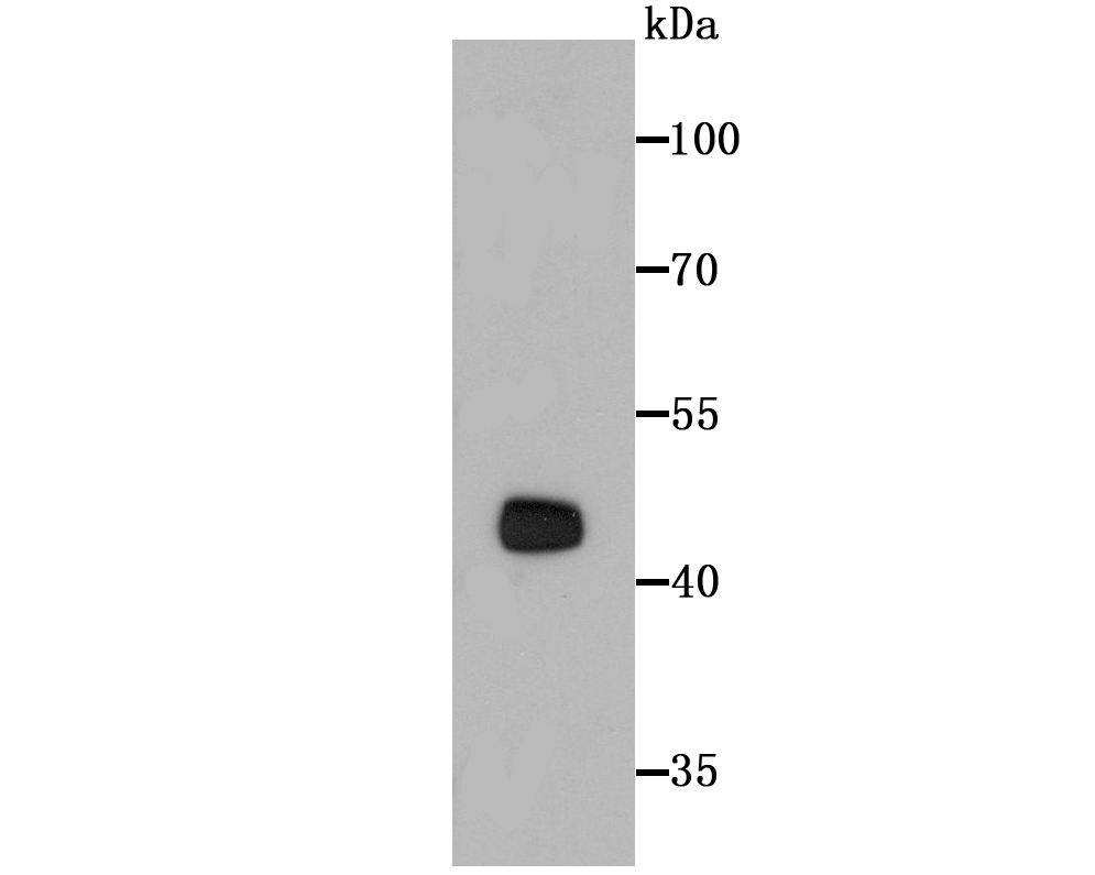
Fig1: Western blot analysis of PTEN on MCF-7 cell lysates using anti-PTEN antibody at 1/500 dilution.Application
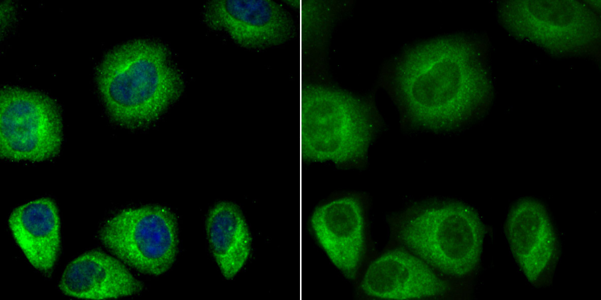
Fig2: ICC staining PTEN in HUVEC cells (green). The nuclear counter stain is DAPI (blue). Cells were fixed in paraformaldehyde, permeabilised with 0.25% Triton X100/PBS.Application
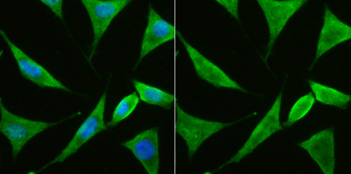
Fig3: ICC staining PTEN in SH-SY5Y cells (green). The nuclear counter stain is DAPI (blue). Cells were fixed in paraformaldehyde, permeabilised with 0.25% Triton X100/PBS.Application
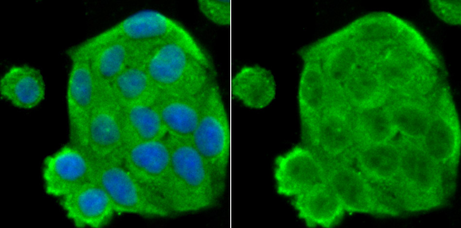
Fig4: ICC staining PTEN in Hela cells (green). The nuclear counter stain is DAPI (blue). Cells were fixed in paraformaldehyde, permeabilised with 0.25% Triton X100/PBS.Application
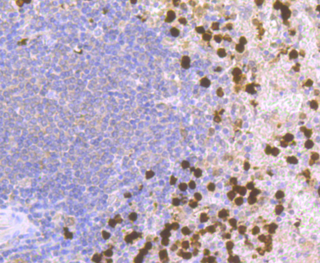
Fig5: Immunohistochemical analysis of paraffin-embedded human spleen tissue using anti-PTEN antibody. Counter stained with hematoxylin.Application
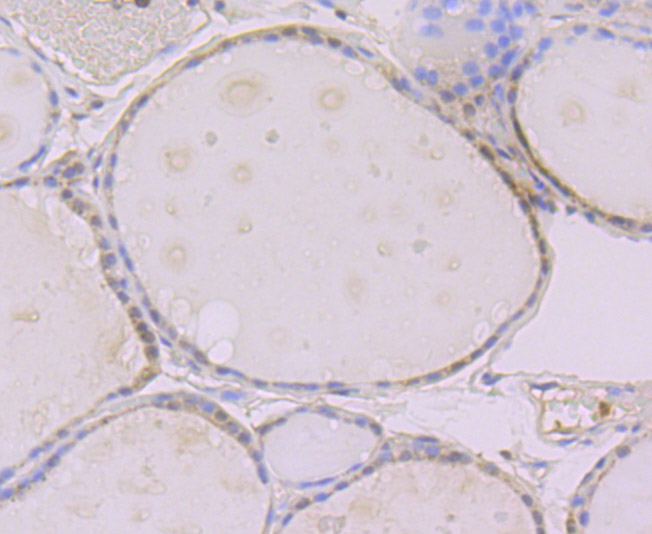
Fig6: Immunohistochemical analysis of paraffin-embedded human thyroid gland tissue using anti-PTEN antibody. Counter stained with hematoxylin.Application
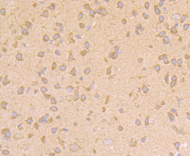
Fig7: Immunohistochemical analysis of paraffin-embedded mouse brain tissue using anti-PTEN antibody. Counter stained with hematoxylin.Application
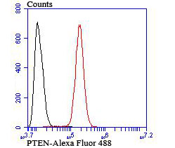
Fig8: Flow cytometric analysis of SH-SY5Y cells with PTEN antibody at 1/100 dilution (red) compared with an unlabelled control (cells without incubation with primary antibody; black).| Product Name | PTEN |
|---|---|
| Antibody Type | Primary Antibodies |
| Antigen Alias | 10q23del antibody BZS antibody DEC antibody GLM2 antibody MGC11227 antibody MHAM antibody MMAC1 antibody MMAC1 phosphatase and tensin homolog deleted on chromosome 10 antibody Mutated in multiple advanced cancers 1 antibody Phosphatase and tensin homolog antibody Phosphatase and tensin like protein antibody Phosphatidylinositol 3,4,5-trisphosphate 3-phosphatase and dual-specificity protein phosphatase PTEN antibody Pten antibody PTEN_HUMAN antibody PTEN1 antibody TEP1 antibody |
| Product description | As human tumors progress to advanced stages, one genetic alteration that occurs at high frequency is a loss of heterozygosity (LOH) at chromosome 10q23. Mapping of homozygous deletions on this chromosome led to the isolation of the PTEN gene, also designated MMAC1 (for mutated in multiple advanced cancers) and TEP1. This candidate tumor suppressor gene exhibits a high frequency of mutations in human glioblastomas and is also mutated in other cancers, including sporadic brain, breast, kidney and prostate cancers. PTEN has been associated with Cowden disease, an autosomal dominant cancer predisposition syndrome. The PTEN gene product is a putative protein tyrosine phosphatase that is localized to the cytoplasm and shares extensive homology with the cytoskeletal proteins tensin and auxilin. Gene transfer studies have indicated that the phosphatase domain of PTEN is essential for growth suppression of glioma cells. |
| Immunogen | Recombinant protein |
| Clonality | Polyclonal |
|---|---|
| Isotype | IgG |
| Host Species | Rabbit |
| Tested Applications | WBICCIHCFC |
| WB:1:500 ICC:1:100-1:500 IHC:1:50-1:200 FC:1:50-1:100 |
|
| Species Reactivity | HumanMouse |
| Concentration | 1mg/ml |
| Alternative Names | 10q23del antibody
BZS antibody
DEC antibody
GLM2 antibody
MGC11227 antibody
MHAM antibody
MMAC1 antibody
MMAC1 phosphatase and tensin homolog deleted on chromosome 10 antibody
Mutated in multiple advanced cancers 1 antibody
Phosphatase and tensin homolog antibody
Phosphatase and tensin like protein antibody
Phosphatidylinositol 3 4 5-trisphosphate 3-phosphatase and dual-specificity protein phosphatase PTEN antibody Pten antibody PTEN_HUMAN antibody PTEN1 antibody TEP1 antibody |
|---|---|
| Molecular Weight(MW) | 47 kDa |
| Cellular Localization | Nucleus. Cytoplasm. Secreted. |
Application
Fig1: Western blot analysis of PTEN on MCF-7 cell lysates using anti-PTEN antibody at 1/500 dilution.
Application
Fig2: ICC staining PTEN in HUVEC cells (green). The nuclear counter stain is DAPI (blue). Cells were fixed in paraformaldehyde, permeabilised with 0.25% Triton X100/PBS.
Application
Fig3: ICC staining PTEN in SH-SY5Y cells (green). The nuclear counter stain is DAPI (blue). Cells were fixed in paraformaldehyde, permeabilised with 0.25% Triton X100/PBS.
Application
Fig4: ICC staining PTEN in Hela cells (green). The nuclear counter stain is DAPI (blue). Cells were fixed in paraformaldehyde, permeabilised with 0.25% Triton X100/PBS.
Application
Fig5: Immunohistochemical analysis of paraffin-embedded human spleen tissue using anti-PTEN antibody. Counter stained with hematoxylin.
Application
Fig6: Immunohistochemical analysis of paraffin-embedded human thyroid gland tissue using anti-PTEN antibody. Counter stained with hematoxylin.
Application
Fig7: Immunohistochemical analysis of paraffin-embedded mouse brain tissue using anti-PTEN antibody. Counter stained with hematoxylin.
Application
Fig8: Flow cytometric analysis of SH-SY5Y cells with PTEN antibody at 1/100 dilution (red) compared with an unlabelled control (cells without incubation with primary antibody; black).| Positive Control | MCF-7, Hela, HUVEC, SH-SY5Y, human thyroid gland tissue, human spleen tissue, mouse brain tissue. |
|---|---|
| Application Notes | WB:1:500 ICC:1:100-1:500 IHC:1:50-1:200 FC:1:50-1:100 |
| Form | Liquid |
|---|---|
| Storage Instructions | Store at +4℃ after thawing. Aliquot store at -20℃ or -80℃. Avoid repeated freeze / thaw cycles. |
| Storage Buffer | 1*TBS (pH7.4), 0.5%BSA, 50%Glycerol. Preservative: 0.05% Sodium Azide. |


