Application
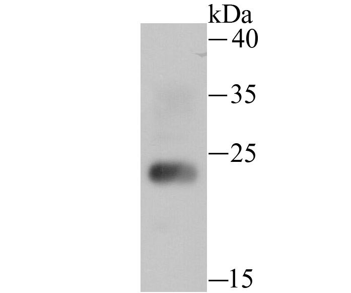
Fig1: Western blot analysis of HMGB2 on mouse testis tissue lysate using anti-HMGB2 antibody at 1/500 dilution.Application
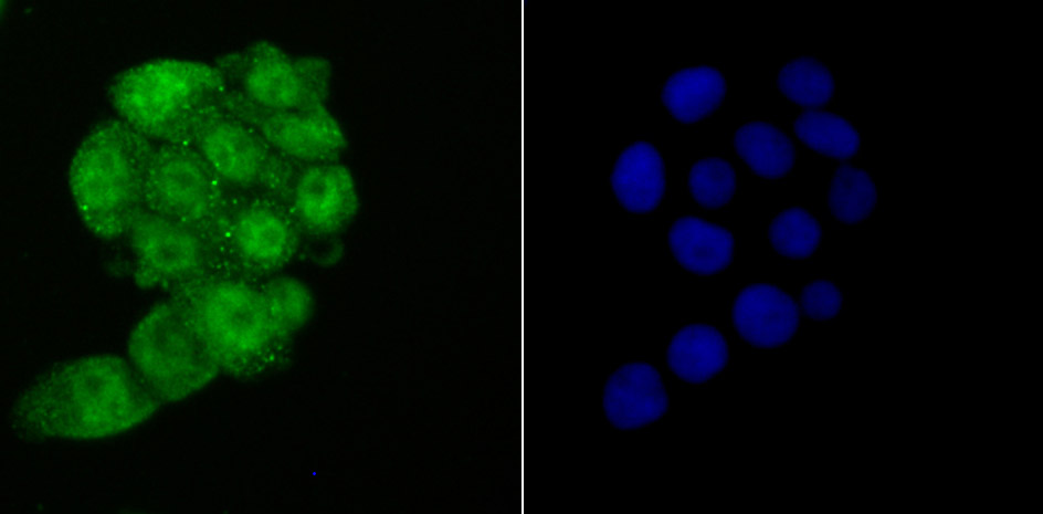
Fig2: ICC staining HMGB2 in LOVO cells (green). The nuclear counter stain is DAPI (blue). Cells were fixed in paraformaldehyde, permeabilised with 0.25% Triton X100/PBS.Application
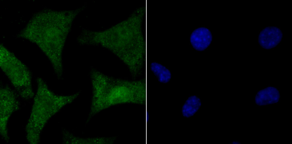
Fig3: ICC staining HMGB2 in SH-SY5Y cells (green). The nuclear counter stain is DAPI (blue). Cells were fixed in paraformaldehyde, permeabilised with 0.25% Triton X100/PBS.Application
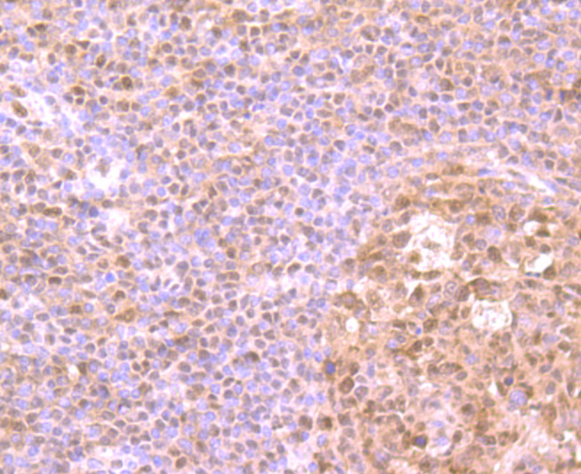
Fig4: Immunohistochemical analysis of paraffin-embedded human tonsil tissue using anti-HMGB2 antibody. Counter stained with hematoxylin.Application
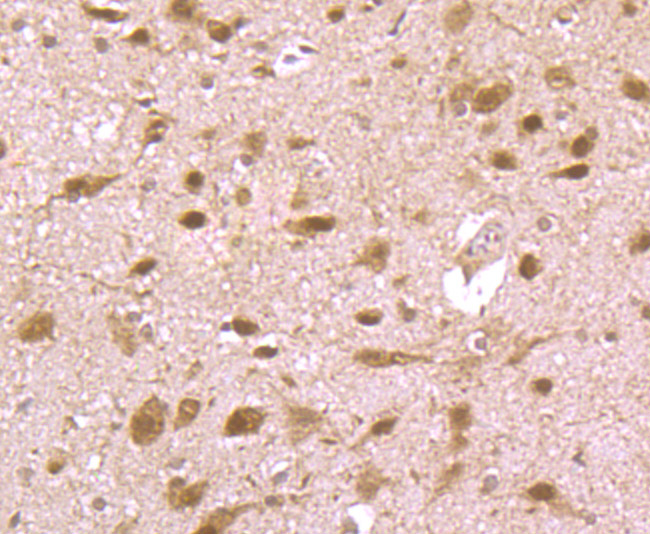
Fig5: Immunohistochemical analysis of paraffin-embedded rat brain tissue using anti-HMGB2 antibody. Counter stained with hematoxylin.Application
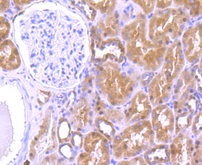
Fig6: Immunohistochemical analysis of paraffin-embedded human kidney tissue using anti-HMGB2 antibody. Counter stained with hematoxylin.Application
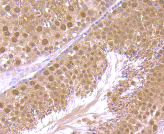
Fig7: Immunohistochemical analysis of paraffin-embedded mouse testis tissue using anti-HMGB2 antibody. Counter stained with hematoxylin.| Product Name | HMGB2 |
|---|---|
| Antibody Type | Primary Antibodies |
| Antigen Alias | C80539 antibody High mobility group (nonhistone chromosomal) protein 2 antibody High mobility group box 2 antibody High mobility group protein 2 antibody High mobility group protein B2 antibody HMG 2 antibody HMG B2 antibody HMG-2 antibody HMG2 antibody HMGB2 antibody HMGB2_HUMAN antibody |
| Product description | High mobility group (HMG) proteins 1 and 2 are ubiquitous non-histone components of chromatin. Evidence suggests that the binding of HMG proteins to DNA induces alterations in the DNA architecture including DNA bending and unwinding of the helix. HMG proteins synergize with Oct-2, members of the NFκB family, ATF-2 and c-Jun to activate transcription. Other studies indicate that phosphorylation of HMG protein is required to stimulate the transcriptional activity of the protein. Human HMG-1 and HMG-2 both contain two DNA-binding domains, termed HMG boxes. HMG proteins bind single-stranded DNA but induce conformational changes in double-stranded DNA alone. |
| Immunogen | Recombinant protein. |
| Clonality | Polyclonal |
|---|---|
| Isotype | IgG |
| Host Species | Rabbit |
| Tested Applications | WBICCIHC |
| WB:1:500-1:1,000 ICC:1:50-1:200 IHC:1:50-1:200 |
|
| Species Reactivity | HumanMouseRat |
| Concentration | 1mg/ml |
| Alternative Names | C80539 antibody High mobility group (nonhistone chromosomal) protein 2 antibody High mobility group box 2 antibody High mobility group protein 2 antibody High mobility group protein B2 antibody HMG 2 antibody HMG B2 antibody HMG-2 antibody HMG2 antibody HMGB2 antibody HMGB2_HUMAN antibody |
|---|---|
| Molecular Weight(MW) | 24 kDa |
| Cellular Localization | Secreted. Nucleus. Cytoplasm. |
Application
Fig1: Western blot analysis of HMGB2 on mouse testis tissue lysate using anti-HMGB2 antibody at 1/500 dilution.
Application
Fig2: ICC staining HMGB2 in LOVO cells (green). The nuclear counter stain is DAPI (blue). Cells were fixed in paraformaldehyde, permeabilised with 0.25% Triton X100/PBS.
Application
Fig3: ICC staining HMGB2 in SH-SY5Y cells (green). The nuclear counter stain is DAPI (blue). Cells were fixed in paraformaldehyde, permeabilised with 0.25% Triton X100/PBS.
Application
Fig4: Immunohistochemical analysis of paraffin-embedded human tonsil tissue using anti-HMGB2 antibody. Counter stained with hematoxylin.
Application
Fig5: Immunohistochemical analysis of paraffin-embedded rat brain tissue using anti-HMGB2 antibody. Counter stained with hematoxylin.
Application
Fig6: Immunohistochemical analysis of paraffin-embedded human kidney tissue using anti-HMGB2 antibody. Counter stained with hematoxylin.
Application
Fig7: Immunohistochemical analysis of paraffin-embedded mouse testis tissue using anti-HMGB2 antibody. Counter stained with hematoxylin.| Positive Control | Mouse testis tissue, LOVO, SH-SY5Y, rat brain tissue, human tonsil tissue, human kidney tissue. |
|---|---|
| Application Notes | WB:1:500-1:1,000 ICC:1:50-1:200 IHC:1:50-1:200 |
| Form | Liquid |
|---|---|
| Storage Instructions | Store at +4℃ after thawing. Aliquot store at -20℃ or -80℃. Avoid repeated freeze / thaw cycles. |
| Storage Buffer | 1*TBS (pH7.4), 0.5%BSA, 50%Glycerol. Preservative: 0.05% Sodium Azide. |


