Application
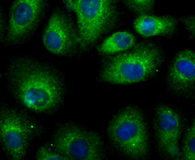
Fig1: ICC staining LRRK2 in A549 cells (green). The nuclear counter stain is DAPI (blue). Cells were fixed in paraformaldehyde, permeabilised with 0.25% Triton X100/PBS.Application
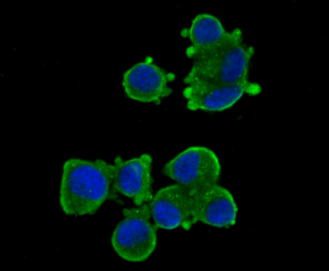
Fig2: ICC staining LRRK2 in N2A cells (green). The nuclear counter stain is DAPI (blue). Cells were fixed in paraformaldehyde, permeabilised with 0.25% Triton X100/PBS.Application
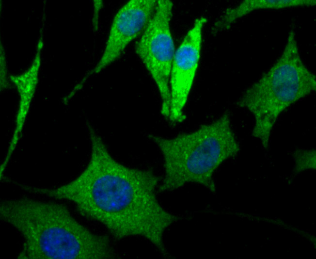
Fig3: ICC staining LRRK2 in SH-SY5Y cells (green). The nuclear counter stain is DAPI (blue). Cells were fixed in paraformaldehyde, permeabilised with 0.25% Triton X100/PBS.Application
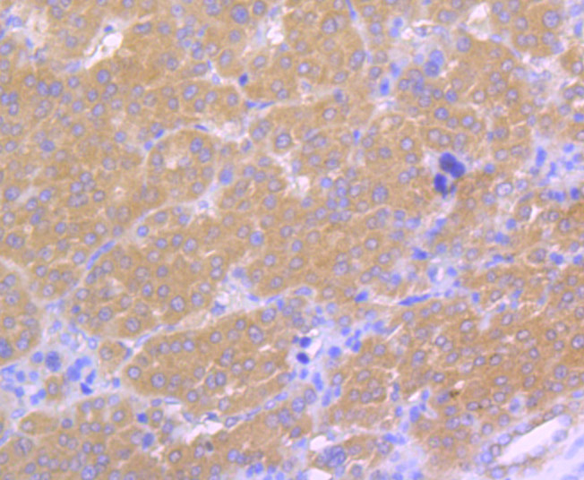
Fig4: Immunohistochemical analysis of paraffin-embedded human liver cancer tissue using anti-LRRK2 antibody. Counter stained with hematoxylin.Application
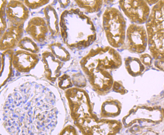
Fig5: Immunohistochemical analysis of paraffin-embedded human kidney tissue using anti-LRRK2 antibody. Counter stained with hematoxylin.Application
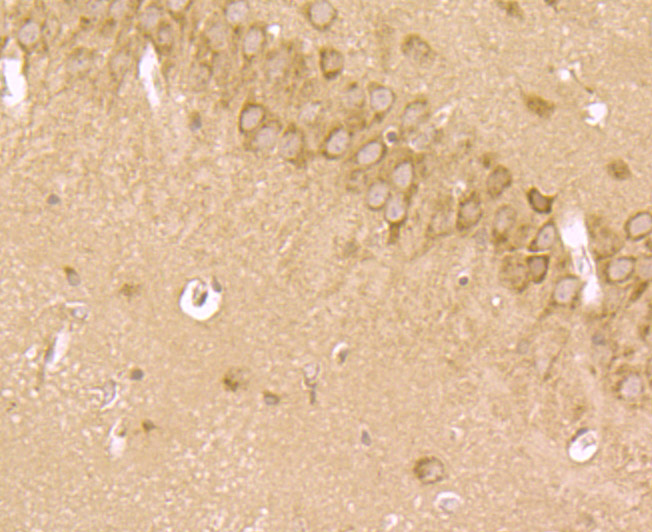
Fig6: Immunohistochemical analysis of paraffin-embedded mouse hippocampus tissue using anti-LRRK2 antibody. Counter stained with hematoxylin.Application
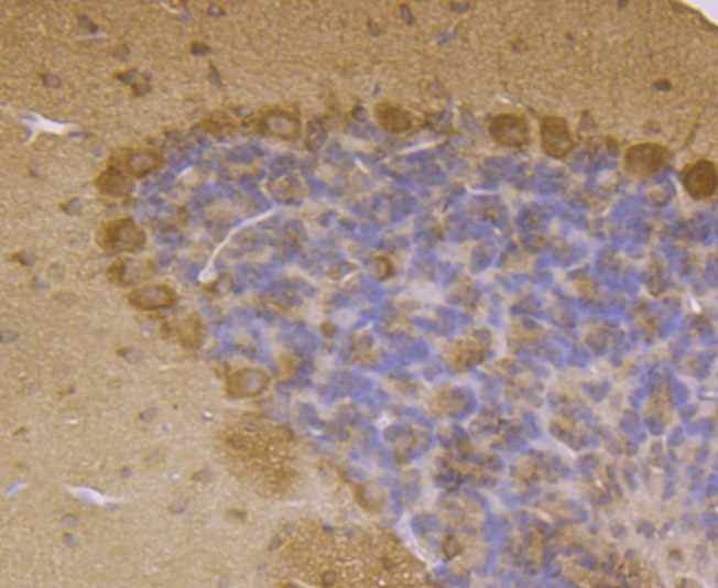
Fig7: Immunohistochemical analysis of paraffin-embedded mouse cerebellum tissue using anti-LRRK2 antibody. Counter stained with hematoxylin.| Product Name | LRRK2 |
|---|---|
| Antibody Type | Primary Antibodies |
| Antigen Alias | augmented in rheumatoid arthritis 17 antibody AURA17 antibody Dardarin antibody Leucine rich repeat kinase 2 antibody leucine rich repeat serine threonine protein kinase 2 antibody Leucine-rich repeat serine/threonine-protein kinase 2 antibody LRRK 2 antibody LRRK2 antibody LRRK2_HUMAN antibody PARK 8 antibody PARK8 antibody RIPK7 antibody ROCO 2 antibody ROCO2 antibody |
| Product description | Parkinson's disease is a disorder of movement, cognition and emotion. It is characterized pathologically by neuronal degeneration with Lewy bodies, which are cytoplasmic inclusion bodies containing deposits of aggregated proteins. Mutations in the leucine-rich repeat kinase 2 gene (LRRK2) cause autosomal-dominant parkinsonism, with clinical features of Parkinson's disease and with pleomorphic pathology including deposits of aggregated protein. The LRRK2 protein consists of multiple domains and belongs to the Roco family, a novel group of the Ras/GTPase superfamily. Besides the GTPase (Roc) domain, it contains a predicted kinase domain, with homology to MAP kinase kinase kinases. LRRK2 is localized in the cytoplasm and is associated with cellular membrane structures. The purified LRRK2 protein demonstrates autokinase activity. |
| Immunogen | Peptide. |
| Clonality | Polyclonal |
|---|---|
| Isotype | IgG |
| Host Species | Rabbit |
| Tested Applications | ICCIHC |
| ICC:1:50-1:200 IHC:1:50-1:200 |
|
| Species Reactivity | HumanMouse |
| Concentration | 1mg/ml |
| Alternative Names | augmented in rheumatoid arthritis 17 antibody AURA17 antibody Dardarin antibody Leucine rich repeat kinase 2 antibody leucine rich repeat serine threonine protein kinase 2 antibody Leucine-rich repeat serine/threonine-protein kinase 2 antibody LRRK 2 antibody LRRK2 antibody LRRK2_HUMAN antibody PARK 8 antibody PARK8 antibody RIPK7 antibody ROCO 2 antibody ROCO2 antibody |
|---|---|
| Molecular Weight(MW) | 286 kDa |
| Cellular Localization | Mitochondrion. Lysosome. Golgi apparatus. |
Application
Fig1: ICC staining LRRK2 in A549 cells (green). The nuclear counter stain is DAPI (blue). Cells were fixed in paraformaldehyde, permeabilised with 0.25% Triton X100/PBS.
Application
Fig2: ICC staining LRRK2 in N2A cells (green). The nuclear counter stain is DAPI (blue). Cells were fixed in paraformaldehyde, permeabilised with 0.25% Triton X100/PBS.
Application
Fig3: ICC staining LRRK2 in SH-SY5Y cells (green). The nuclear counter stain is DAPI (blue). Cells were fixed in paraformaldehyde, permeabilised with 0.25% Triton X100/PBS.
Application
Fig4: Immunohistochemical analysis of paraffin-embedded human liver cancer tissue using anti-LRRK2 antibody. Counter stained with hematoxylin.
Application
Fig5: Immunohistochemical analysis of paraffin-embedded human kidney tissue using anti-LRRK2 antibody. Counter stained with hematoxylin.
Application
Fig6: Immunohistochemical analysis of paraffin-embedded mouse hippocampus tissue using anti-LRRK2 antibody. Counter stained with hematoxylin.
Application
Fig7: Immunohistochemical analysis of paraffin-embedded mouse cerebellum tissue using anti-LRRK2 antibody. Counter stained with hematoxylin.| Positive Control | HepG2, N2A, SH-SY5Y, human liver cancer tissue, human kidney tissue, mouse hippocampus tissue, mouse cerebellum tissue. |
|---|---|
| Application Notes | ICC:1:50-1:200 IHC:1:50-1:200 |
| Form | Liquid |
|---|---|
| Storage Instructions | Store at +4℃ after thawing. Aliquot store at -20℃ or -80℃. Avoid repeated freeze / thaw cycles. |
| Storage Buffer | 1*TBS (pH7.4), 0.5%BSA, 50%Glycerol. Preservative: 0.05% Sodium Azide. |


