Application
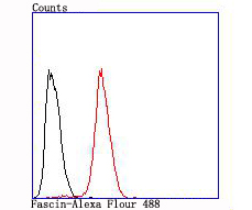
Fig8: Flow cytometric analysis of Hela cells with Fascin antibody at 1/100 dilution (red) compared with an unlabelled control (cells without incubation with primary antibody; black).Application
Fig8: Flow cytometric analysis of Hela cells with Fascin antibody at 1/100 dilution (red) compared with an unlabelled control (cells without incubation with primary antibody; black).Application
Fig8: Flow cytometric analysis of Hela cells with Fascin antibody at 1/100 dilution (red) compared with an unlabelled control (cells without incubation with primary antibody; black).Application
Fig8: Flow cytometric analysis of Hela cells with Fascin antibody at 1/100 dilution (red) compared with an unlabelled control (cells without incubation with primary antibody; black).Application
Fig8: Flow cytometric analysis of Hela cells with Fascin antibody at 1/100 dilution (red) compared with an unlabelled control (cells without incubation with primary antibody; black).Application
Fig8: Flow cytometric analysis of Hela cells with Fascin antibody at 1/100 dilution (red) compared with an unlabelled control (cells without incubation with primary antibody; black).Application
Fig8: Flow cytometric analysis of Hela cells with Fascin antibody at 1/100 dilution (red) compared with an unlabelled control (cells without incubation with primary antibody; black).Application
Fig8: Flow cytometric analysis of Hela cells with Fascin antibody at 1/100 dilution (red) compared with an unlabelled control (cells without incubation with primary antibody; black).| Product Name | Fascin [JM12-53] |
|---|---|
| Antibody Type | Primary Antibodies |
| Antigen Alias | 55 kDa actin bundling protein antibody 55 kDa actin-bundling protein antibody Actin bundling protein antibody actin bundling protein, 55-KD antibody FAN 1 antibody FAN1 antibody Fascin 1 antibody Fascin actin bundling protein 1 antibody Fascin antibody Fascin homolog 1 actin bundling protein (Strongylocentrotus purpuratus) antibody Fascin homolog 1 antibody Fascin, sea urchin, homolog of, 1 antibody Fascin1 antibody FLJ38511 antibody FSCN 1 antibody FSCN1 antibody FSCN1_HUMAN antibody HSN antibody p55 antibody Singed (Drosophila) like (sea urchin fascin homolog like) antibody Singed drosophila homolog like antibody Singed like (fascin homolog sea urchin) (Drosophila) antibody Singed like (fascin homolog sea urchin) antibody Singed like protein antibody Singed, drosophila, homolog of antibody Singed-like protein antibody SNL antibody Strongylocentrotus purpuratus antibody |
| Product description | Cell adhesion to extracellular matrix is an important physiological stimulus for organization of the Actin-based cytoskeleton. Adhesion to the matrix glycoprotein Thrombospondin 1 triggers the sustained formation of F-Actin microspikes that contain the Actin-bundling protein Fascin. These structures are also implicated in cell migration, which may be an important function of Thrombospondin 1 in tissue remodelling and wound repair. Fascin bundles Actin microfilaments within dynamic cellular structures such as microspikes, stress fibers and membrane ruffles. Fascin could serve as a prognostic factor for abnormal ovarian epithelial pathology and could be a novel target for the treatment of ovarian cancer. Fascin, an Actin-bundling protein, identifies dendritic cells in the blood and in tissues. |
| Immunogen | recombinant protein |
| Clonality | Monoclonal |
|---|---|
| Isotype | IgG |
| Host Species | Recombinant rabbit |
| Tested Applications | WBICC/IFIHCIPFC |
| WB:1:500-1:1,000 ICC:1:50-1:200 IHC:1:50-1:200 FC:1:50-1:100 |
|
| Species Reactivity | HumanMouseRat |
| Concentration | 1mg/ml |
| Alternative Names | 55 kDa actin bundling protein antibody
55 kDa actin-bundling protein antibody
Actin bundling protein antibody
actin bundling protein 55-KD antibody FAN 1 antibody FAN1 antibody Fascin 1 antibody Fascin actin bundling protein 1 antibody Fascin antibody Fascin homolog 1 actin bundling protein (Strongylocentrotus purpuratus) antibody Fascin homolog 1 antibody Fascin sea urchin homolog of 1 antibody Fascin1 antibody FLJ38511 antibody FSCN 1 antibody FSCN1 antibody FSCN1_HUMAN antibody HSN antibody p55 antibody Singed (Drosophila) like (sea urchin fascin homolog like) antibody Singed drosophila homolog like antibody Singed like (fascin homolog sea urchin) (Drosophila) antibody Singed like (fascin homolog sea urchin) antibody Singed like protein antibody Singed drosophila homolog of antibody Singed-like protein antibody SNL antibody Strongylocentrotus purpuratus antibody |
|---|---|
| Molecular Weight(MW) | 54 kDa |
| Cellular Localization | Cytoplasm. |
Application
Fig8: Flow cytometric analysis of Hela cells with Fascin antibody at 1/100 dilution (red) compared with an unlabelled control (cells without incubation with primary antibody; black).
Application
Fig8: Flow cytometric analysis of Hela cells with Fascin antibody at 1/100 dilution (red) compared with an unlabelled control (cells without incubation with primary antibody; black).
Application
Fig8: Flow cytometric analysis of Hela cells with Fascin antibody at 1/100 dilution (red) compared with an unlabelled control (cells without incubation with primary antibody; black).
Application
Fig8: Flow cytometric analysis of Hela cells with Fascin antibody at 1/100 dilution (red) compared with an unlabelled control (cells without incubation with primary antibody; black).
Application
Fig8: Flow cytometric analysis of Hela cells with Fascin antibody at 1/100 dilution (red) compared with an unlabelled control (cells without incubation with primary antibody; black).
Application
Fig8: Flow cytometric analysis of Hela cells with Fascin antibody at 1/100 dilution (red) compared with an unlabelled control (cells without incubation with primary antibody; black).
Application
Fig8: Flow cytometric analysis of Hela cells with Fascin antibody at 1/100 dilution (red) compared with an unlabelled control (cells without incubation with primary antibody; black).
Application
Fig8: Flow cytometric analysis of Hela cells with Fascin antibody at 1/100 dilution (red) compared with an unlabelled control (cells without incubation with primary antibody; black).| Positive Control | SH-SY5Y, human brain tissue lysate, rat spleen tissue lysate, mouse testes tissue lysate, mouse brain tissue lysate, mouse spleen tissue lysate, Hela, SW480, rat brain tissue, human tonsil tissue, human kidney tissue. |
|---|---|
| Application Notes | WB:1:500-1:1,000 ICC:1:50-1:200 IHC:1:50-1:200 FC:1:50-1:100 |
| Form | Liquid |
|---|---|
| Storage Instructions | Store at +4℃ after thawing. Aliquot store at -20℃ or -80℃. Avoid repeated freeze / thaw cycles. |
| Storage Buffer | 1*TBS (pH7.4), 1%BSA, 40%Glycerol. Preservative: 0.05% Sodium Azide. |


