Application
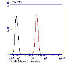
Fig7: Flow cytometric analysis of Jurkat cells with IL4 antibody at 1/100 dilution (red) compared with an unlabelled control (cells without incubation with primary antibody; black). Alexa Fluor 488-conjugated goat anti rabbit IgG was used as the secondary antibody.Application
Fig7: Flow cytometric analysis of Jurkat cells with IL4 antibody at 1/100 dilution (red) compared with an unlabelled control (cells without incubation with primary antibody; black). Alexa Fluor 488-conjugated goat anti rabbit IgG was used as the secondary antibody.Application
Fig7: Flow cytometric analysis of Jurkat cells with IL4 antibody at 1/100 dilution (red) compared with an unlabelled control (cells without incubation with primary antibody; black). Alexa Fluor 488-conjugated goat anti rabbit IgG was used as the secondary antibody.Application
Fig7: Flow cytometric analysis of Jurkat cells with IL4 antibody at 1/100 dilution (red) compared with an unlabelled control (cells without incubation with primary antibody; black). Alexa Fluor 488-conjugated goat anti rabbit IgG was used as the secondary antibody.Application
Fig7: Flow cytometric analysis of Jurkat cells with IL4 antibody at 1/100 dilution (red) compared with an unlabelled control (cells without incubation with primary antibody; black). Alexa Fluor 488-conjugated goat anti rabbit IgG was used as the secondary antibody.Application
Fig7: Flow cytometric analysis of Jurkat cells with IL4 antibody at 1/100 dilution (red) compared with an unlabelled control (cells without incubation with primary antibody; black). Alexa Fluor 488-conjugated goat anti rabbit IgG was used as the secondary antibody.Application
Fig7: Flow cytometric analysis of Jurkat cells with IL4 antibody at 1/100 dilution (red) compared with an unlabelled control (cells without incubation with primary antibody; black). Alexa Fluor 488-conjugated goat anti rabbit IgG was used as the secondary antibody.| Product Name | IL4 |
|---|---|
| Antibody Type | Primary Antibodies |
| Antigen Alias | B cell growth factor 1 antibody B cell IgG differentiation factor antibody B Cell Stimulatory Factor 1 antibody B-cell stimulatory factor 1 antibody BCGF 1 antibody BCGF1 antibody Binetrakin antibody BSF-1 antibody BSF1 antibody IGG1 induction factor antibody IL 4 antibody IL-4 antibody IL4 antibody IL4_HUMAN antibody Il4e12 antibody Interleukin 4 antibody Interleukin 4 variant 2 antibody Interleukin 4, isoform 1 antibody Interleukin-4 antibody Lymphocyte stimulatory factor 1 antibody MGC79402 antibody Pitrakinra antibody |
| Product description | Interleukin-4 (IL-4), also designated B cell stimulatory factor-1, is a glycosylated cytokine secreted by activated T lymphocytes, basophils and mast cells. The secreted IL-4 protein promotes the growth and differentiation of cells that participate in immune defense by favoring such events as the expansion of the Th2 lineage relative to Th1 cells. These T helper cell subsets are defined by their pattern of cytokine secretion: Th1 cells secrete IL-2, TNFβ and IFN-γ, while Th2 cells secrete IL-4, IL-5 and IL-10. Another key immunological function of IL-4 is to induce immunoglobulin class switching. IL-4 has been shown to induce the production of IgE and enhance IgG4 secretion by B cells, but suppress the production of IgM, IgA, IgG1, IgG2, and IgG3. It has been determined that Stat6 is rapidly tyrosine phosphorylated following stimulation of IL-3 or IL-4, but is not detectably phosphorylated following stimulation with IL-2, IL-12 or erythropoietin. |
| Immunogen | Recombinant protein. |
| Clonality | Polyclonal |
|---|---|
| Isotype | IgG |
| Host Species | Rabbit |
| Tested Applications | WBICCIHCFC |
| WB:1:500-1:1,000 ICC:1:50-1:200 IHC:1:50-1:200 FC:1:50-1:100 |
|
| Species Reactivity | HumanMouse |
| Concentration | 1mg/ml |
| Alternative Names | B cell growth factor 1 antibody
B cell IgG differentiation factor antibody
B Cell Stimulatory Factor 1 antibody
B-cell stimulatory factor 1 antibody
BCGF 1 antibody
BCGF1 antibody
Binetrakin antibody
BSF-1 antibody
BSF1 antibody
IGG1 induction factor antibody
IL 4 antibody
IL-4 antibody
IL4 antibody
IL4_HUMAN antibody
Il4e12 antibody
Interleukin 4 antibody
Interleukin 4 variant 2 antibody
Interleukin 4 isoform 1 antibody Interleukin-4 antibody Lymphocyte stimulatory factor 1 antibody MGC79402 antibody Pitrakinra antibody |
|---|---|
| Molecular Weight(MW) | 17 kDa |
| Cellular Localization | Secreted. |
Application
Fig7: Flow cytometric analysis of Jurkat cells with IL4 antibody at 1/100 dilution (red) compared with an unlabelled control (cells without incubation with primary antibody; black). Alexa Fluor 488-conjugated goat anti rabbit IgG was used as the secondary antibody.
Application
Fig7: Flow cytometric analysis of Jurkat cells with IL4 antibody at 1/100 dilution (red) compared with an unlabelled control (cells without incubation with primary antibody; black). Alexa Fluor 488-conjugated goat anti rabbit IgG was used as the secondary antibody.
Application
Fig7: Flow cytometric analysis of Jurkat cells with IL4 antibody at 1/100 dilution (red) compared with an unlabelled control (cells without incubation with primary antibody; black). Alexa Fluor 488-conjugated goat anti rabbit IgG was used as the secondary antibody.
Application
Fig7: Flow cytometric analysis of Jurkat cells with IL4 antibody at 1/100 dilution (red) compared with an unlabelled control (cells without incubation with primary antibody; black). Alexa Fluor 488-conjugated goat anti rabbit IgG was used as the secondary antibody.
Application
Fig7: Flow cytometric analysis of Jurkat cells with IL4 antibody at 1/100 dilution (red) compared with an unlabelled control (cells without incubation with primary antibody; black). Alexa Fluor 488-conjugated goat anti rabbit IgG was used as the secondary antibody.
Application
Fig7: Flow cytometric analysis of Jurkat cells with IL4 antibody at 1/100 dilution (red) compared with an unlabelled control (cells without incubation with primary antibody; black). Alexa Fluor 488-conjugated goat anti rabbit IgG was used as the secondary antibody.
Application
Fig7: Flow cytometric analysis of Jurkat cells with IL4 antibody at 1/100 dilution (red) compared with an unlabelled control (cells without incubation with primary antibody; black). Alexa Fluor 488-conjugated goat anti rabbit IgG was used as the secondary antibody.| Positive Control | Recombinant protein lysate, human spleen tissue, human breast cancer tissue, mouse colon tissue, mouse small intestine tissue, Jurkat. |
|---|---|
| Application Notes | WB:1:500-1:1,000 ICC:1:50-1:200 IHC:1:50-1:200 FC:1:50-1:100 |
| Form | Liquid |
|---|---|
| Storage Instructions | Store at +4℃ after thawing. Aliquot store at -20℃ or -80℃. Avoid repeated freeze / thaw cycles. |
| Storage Buffer | 1*TBS (pH7.4), 0.5%BSA, 50%Glycerol. Preservative: 0.05% Sodium Azide. |


