Application
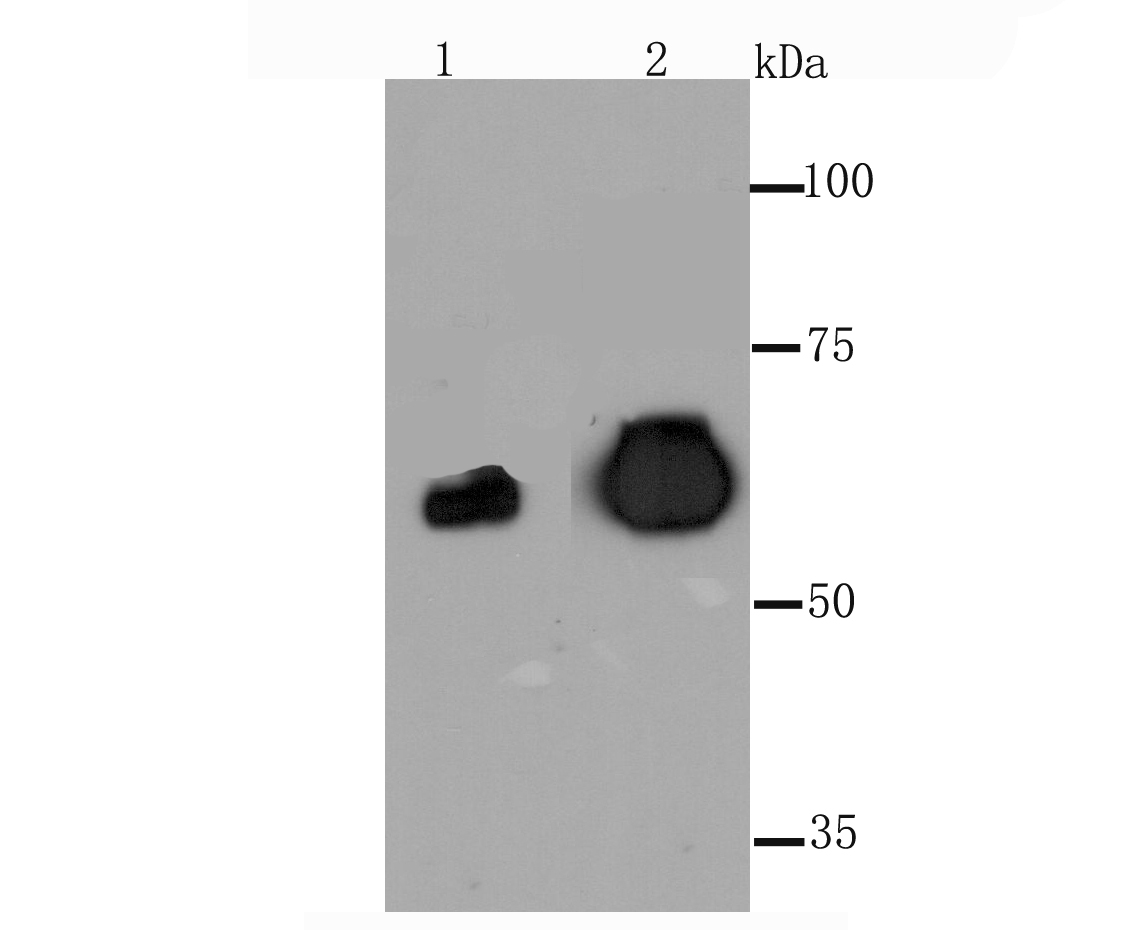
Fig1: Western blot analysis of PLAP on human placenta tissue (1) and Hela cell (2) lysate using anti-PLAP antibody at 1/1,000 dilution.Application
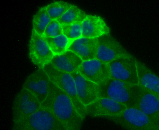
Fig2: ICC staining PLAP in Hela cells (green). The nuclear counter stain is DAPI (blue). Cells were fixed in paraformaldehyde, permeabilised with 0.25% Triton X100/PBS.Application
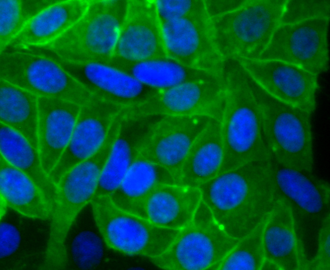
Fig3: ICC staining PLAP in HepG2 cells (green). The nuclear counter stain is DAPI (blue). Cells were fixed in paraformaldehyde, permeabilised with 0.25% Triton X100/PBS.Application
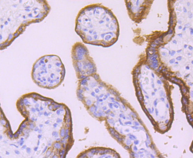
Fig4: Immunohistochemical analysis of paraffin-embedded human placenta tissue using anti-PLAP antibody. Counter stained with hematoxylin.| Product Name | Placental alkaline phosphatase [JM22-53] |
|---|---|
| Antibody Type | Primary Antibodies |
| Antigen Alias | Alkaline phosphatase antibody Alkaline phosphatase placental antibody Alkaline phosphatase placental type antibody Alkaline phosphatase Regan isozyme antibody ALP antibody Alp1 antibody ALPP antibody FLJ61142 antibody Germ-cell alkaline phosphatase antibody nagao isozyme antibody OTTHUMP00000164354 antibody PALP antibody Placental alkaline phosphatase 1 antibody placental heat-stable alkaline phosphatase antibody placental type antibody PLAP antibody PLAP-1 antibody PLAP1 antibody PPB1_HUMAN antibody |
| Product description | Alkaline phosphatases (AP) are glycosyl-phosphatidylinositol (GPI)-anchored, dimeric, Zn2+-metallated glycoproteins that catalyze the hydrolysis of phosphomonoesters into an inorganic phosphate and an alcohol. Placental alkaline phosphatase (also known as PLAP, ALPP, PALP, placental ALP-1 or Regan isozyme) is a 530 amino acid, tissue-specific AP that is expressed in the placenta, the serum of pregnant women and ectopically expressed in various cancers, including those of the ovary and testis. PLAP may assist in guiding migratory cells and transporting specific molecules, such as fatty acids and immunoglobulins, across the plasma membrane. The three tissue-specific APs identified in human, PLAP, germ cell AP (GCAP) and intestinal AP, are 90-98% homologous and their genes are clustered on chromosome 2q. |
| Immunogen | recombinant protein |
| Clonality | Monoclonal |
|---|---|
| Isotype | IgG |
| Host Species | Recombinant rabbit |
| Tested Applications | WBICC/IFIHC |
| WB:1:500-1:2,000 ICC:1:50-1:200 IHC:1:50-1:200 |
|
| Species Reactivity | Human |
| Concentration | 1mg/ml |
| Alternative Names | Alkaline phosphatase antibody Alkaline phosphatase placental antibody Alkaline phosphatase placental type antibody Alkaline phosphatase Regan isozyme antibody ALP antibody Alp1 antibody ALPP antibody FLJ61142 antibody Germ-cell alkaline phosphatase antibody nagao isozyme antibody OTTHUMP00000164354 antibody PALP antibody Placental alkaline phosphatase 1 antibody placental heat-stable alkaline phosphatase antibody placental type antibody PLAP antibody PLAP-1 antibody PLAP1 antibody PPB1_HUMAN antibody |
|---|---|
| Molecular Weight(MW) | 70 kDa |
| Cellular Localization | Cell membrane. |
| SwissProt ID | P05187 |
|---|
Application
Fig1: Western blot analysis of PLAP on human placenta tissue (1) and Hela cell (2) lysate using anti-PLAP antibody at 1/1,000 dilution.
Application
Fig2: ICC staining PLAP in Hela cells (green). The nuclear counter stain is DAPI (blue). Cells were fixed in paraformaldehyde, permeabilised with 0.25% Triton X100/PBS.
Application
Fig3: ICC staining PLAP in HepG2 cells (green). The nuclear counter stain is DAPI (blue). Cells were fixed in paraformaldehyde, permeabilised with 0.25% Triton X100/PBS.
Application
Fig4: Immunohistochemical analysis of paraffin-embedded human placenta tissue using anti-PLAP antibody. Counter stained with hematoxylin.| Positive Control | Human placenta tissue, Hela. |
|---|---|
| Application Notes | WB:1:500-1:2,000 ICC:1:50-1:200 IHC:1:50-1:200 |
| Form | Liquid |
|---|---|
| Storage Instructions | Store at +4℃ after thawing. Aliquot store at -20℃ or -80℃. Avoid repeated freeze / thaw cycles. |
| Storage Buffer | 1*TBS (pH7.4), 1%BSA, 40%Glycerol. Preservative: 0.05% Sodium Azide. |


