Application
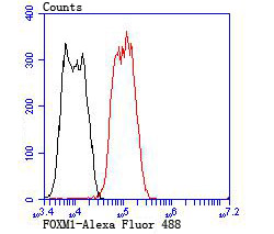
Fig4: Flow cytometric analysis of LOVO cells with FOXM1 antibody at 1/100 dilution (red) compared with an unlabelled control (cells without incubation with primary antibody; black). Alexa Fluor 488-conjugated goat anti rabbit IgG was used as the secondary antibody.Application
Fig4: Flow cytometric analysis of LOVO cells with FOXM1 antibody at 1/100 dilution (red) compared with an unlabelled control (cells without incubation with primary antibody; black). Alexa Fluor 488-conjugated goat anti rabbit IgG was used as the secondary antibody.Application
Fig4: Flow cytometric analysis of LOVO cells with FOXM1 antibody at 1/100 dilution (red) compared with an unlabelled control (cells without incubation with primary antibody; black). Alexa Fluor 488-conjugated goat anti rabbit IgG was used as the secondary antibody.Application
Fig4: Flow cytometric analysis of LOVO cells with FOXM1 antibody at 1/100 dilution (red) compared with an unlabelled control (cells without incubation with primary antibody; black). Alexa Fluor 488-conjugated goat anti rabbit IgG was used as the secondary antibody.| Product Name | FOXM1 |
|---|---|
| Antibody Type | Primary Antibodies |
| Antigen Alias | FKHL16 antibody Forkhead box M1 antibody Forkhead box protein M1 antibody forkhead like 16 antibody Forkhead-related protein FKHL16 antibody FOX M1 antibody Foxm1 antibody FOXM1_HUMAN antibody FOXM1B antibody Hepatocyte nuclear factor 3 forkhead homolog 11 antibody HFH-11 antibody HFH11 antibody HNF-3/fork-head homolog 11 antibody HNF3 antibody INS1 antibody M phase phosphoprotein 2 antibody M-phase phosphoprotein 2 antibody MPHOSPH2 antibody MPM-2 reactive phosphoprotein 2 antibody MPP2 antibody PIG29 antibody TGT3 antibody Transcription factor Trident antibody Trident antibody WIN antibody Winged-helix factor from INS-1 cells antibody Winged-helix factor from INS1 cells antibody |
| Product description | The Fox family of transcription factors is a large group of proteins that share a common DNA binding domain termed a winged-helix or forkhead domain. FOXM1, also known as FKHL16, MPP2 or Trident, is primarily expressed in proliferating cells. The gene encoding human FOXM1 maps to chromosome 12p13. The transcription element that restricts FOXM1 expression to proliferating cells is located 300 bp upstream of the start codon. FOXM1 is most abundant in thymus, testis, small intestine and colon. Alternative splicing generates FOXM1A and FOXM1B isoforms that contain PEST regions involved in rapid protein degradation. A decrease in FOXM1 expression is associated with age-related defects in cellular proliferation. Conversely, an increase in FOXM1B expression in the livers of older transgenic mice restore hepatocyte DNA replication rates to the higher rate present in young livers. FOXM1B activates the transcription of cyclin B1, cyclin D1 and Cdc25B. |
| Immunogen | Recombinant protein. |
| Clonality | Polyclonal |
|---|---|
| Isotype | IgG |
| Host Species | Rabbit |
| Tested Applications | WBICCFC |
| ICC:1:500-1:2,000 FC:1:50-1:100 WB:1:500 |
|
| Species Reactivity | HumanMouse |
| Concentration | 1mg/ml |
| Alternative Names | FKHL16 antibody Forkhead box M1 antibody Forkhead box protein M1 antibody forkhead like 16 antibody Forkhead-related protein FKHL16 antibody FOX M1 antibody Foxm1 antibody FOXM1_HUMAN antibody FOXM1B antibody Hepatocyte nuclear factor 3 forkhead homolog 11 antibody HFH-11 antibody HFH11 antibody HNF-3/fork-head homolog 11 antibody HNF3 antibody INS1 antibody M phase phosphoprotein 2 antibody M-phase phosphoprotein 2 antibody MPHOSPH2 antibody MPM-2 reactive phosphoprotein 2 antibody MPP2 antibody PIG29 antibody TGT3 antibody Transcription factor Trident antibody Trident antibody WIN antibody Winged-helix factor from INS-1 cells antibody Winged-helix factor from INS1 cells antibody |
|---|---|
| Molecular Weight(MW) | 84 kDa |
| Cellular Localization | Nucleus. |
| SwissProt ID | Q08050 |
|---|
Application
Fig4: Flow cytometric analysis of LOVO cells with FOXM1 antibody at 1/100 dilution (red) compared with an unlabelled control (cells without incubation with primary antibody; black). Alexa Fluor 488-conjugated goat anti rabbit IgG was used as the secondary antibody.
Application
Fig4: Flow cytometric analysis of LOVO cells with FOXM1 antibody at 1/100 dilution (red) compared with an unlabelled control (cells without incubation with primary antibody; black). Alexa Fluor 488-conjugated goat anti rabbit IgG was used as the secondary antibody.
Application
Fig4: Flow cytometric analysis of LOVO cells with FOXM1 antibody at 1/100 dilution (red) compared with an unlabelled control (cells without incubation with primary antibody; black). Alexa Fluor 488-conjugated goat anti rabbit IgG was used as the secondary antibody.
Application
Fig4: Flow cytometric analysis of LOVO cells with FOXM1 antibody at 1/100 dilution (red) compared with an unlabelled control (cells without incubation with primary antibody; black). Alexa Fluor 488-conjugated goat anti rabbit IgG was used as the secondary antibody.| Positive Control | A549, HUVEC, LOVO. |
|---|---|
| Application Notes | ICC:1:500-1:2,000 FC:1:50-1:100 WB:1:500 |
| Form | Liquid |
|---|---|
| Storage Instructions | Store at +4℃ after thawing. Aliquot store at -20℃ or -80℃. Avoid repeated freeze / thaw cycles. |
| Storage Buffer | 1*TBS (pH7.4), 0.5%BSA, 50%Glycerol. Preservative: 0.05% Sodium Azide. |


