Application
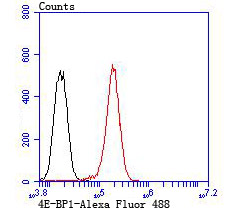
Fig7: Flow cytometric analysis of PC-3M cells with 4E-BP1 antibody at 1/100 dilution (red) compared with an unlabelled control (cells without incubation with primary antibody; black). Alexa Fluor 488-conjugated goat anti rabbit IgG was used as the secondary antibody.Application
Fig7: Flow cytometric analysis of PC-3M cells with 4E-BP1 antibody at 1/100 dilution (red) compared with an unlabelled control (cells without incubation with primary antibody; black). Alexa Fluor 488-conjugated goat anti rabbit IgG was used as the secondary antibody.Application
Fig7: Flow cytometric analysis of PC-3M cells with 4E-BP1 antibody at 1/100 dilution (red) compared with an unlabelled control (cells without incubation with primary antibody; black). Alexa Fluor 488-conjugated goat anti rabbit IgG was used as the secondary antibody.Application
Fig7: Flow cytometric analysis of PC-3M cells with 4E-BP1 antibody at 1/100 dilution (red) compared with an unlabelled control (cells without incubation with primary antibody; black). Alexa Fluor 488-conjugated goat anti rabbit IgG was used as the secondary antibody.Application
Fig7: Flow cytometric analysis of PC-3M cells with 4E-BP1 antibody at 1/100 dilution (red) compared with an unlabelled control (cells without incubation with primary antibody; black). Alexa Fluor 488-conjugated goat anti rabbit IgG was used as the secondary antibody.Application
Fig7: Flow cytometric analysis of PC-3M cells with 4E-BP1 antibody at 1/100 dilution (red) compared with an unlabelled control (cells without incubation with primary antibody; black). Alexa Fluor 488-conjugated goat anti rabbit IgG was used as the secondary antibody.Application
Fig7: Flow cytometric analysis of PC-3M cells with 4E-BP1 antibody at 1/100 dilution (red) compared with an unlabelled control (cells without incubation with primary antibody; black). Alexa Fluor 488-conjugated goat anti rabbit IgG was used as the secondary antibody.| Product Name | 4E-BP1 |
|---|---|
| Antibody Type | Primary Antibodies |
| Antigen Alias | 4E-BP1 antibody 4EBP1 antibody 4EBP1_HUMAN antibody BP 1 antibody eIF4E binding protein 1 antibody eIF4E-binding protein 1 antibody Eif4ebp1 antibody Eukaryotic translation initiation factor 4E-binding protein 1 antibody PHAS-I antibody PHASI antibody Phosphorylated heat- and acid-stable protein regulated by insulin 1 antibody |
| Product description | The translation of proteins from eukaryotic mRNA is initiated by the multisubunit complex eIF-4F, which associates with the mRNA 5' cap structure. eIF-4E, a component of eIF-4F, is responsible for binding to the 5' cap structure and for the assembly of the eIF-4F complex. The regulatory protein 4E-BP1, also referred to as PHAS-I, inhibits eIF-4E function. Phosphorylation of 4E-BP1 by S6 kinase p70, MAP kinases or PKCs causes the disassociation of 4E-BP1 from eIF-4E, promoting translation. A protein that is functionally related to 4E-BP1, designated 4E-BP2, also associates with eIF-4E. |
| Immunogen | Recombinant protein. |
| Clonality | Polyclonal |
|---|---|
| Isotype | IgG |
| Host Species | Rabbit |
| Tested Applications | WBICCIHCFC |
| WB:1:500-1:1,000 ICC:1:500-1:1,000 IHC:1:50-1:200 FC:1:50-1:100 |
|
| Species Reactivity | HumanMouseRat |
| Concentration | 1mg/ml |
| Alternative Names | 4E-BP1 antibody 4EBP1 antibody 4EBP1_HUMAN antibody BP 1 antibody eIF4E binding protein 1 antibody eIF4E-binding protein 1 antibody Eif4ebp1 antibody Eukaryotic translation initiation factor 4E-binding protein 1 antibody PHAS-I antibody PHASI antibody Phosphorylated heat- and acid-stable protein regulated by insulin 1 antibody |
|---|---|
| Molecular Weight(MW) | 12.5 kDa |
| Cellular Localization | Cytoplasm. Nucleus. |
Application
Fig7: Flow cytometric analysis of PC-3M cells with 4E-BP1 antibody at 1/100 dilution (red) compared with an unlabelled control (cells without incubation with primary antibody; black). Alexa Fluor 488-conjugated goat anti rabbit IgG was used as the secondary antibody.
Application
Fig7: Flow cytometric analysis of PC-3M cells with 4E-BP1 antibody at 1/100 dilution (red) compared with an unlabelled control (cells without incubation with primary antibody; black). Alexa Fluor 488-conjugated goat anti rabbit IgG was used as the secondary antibody.
Application
Fig7: Flow cytometric analysis of PC-3M cells with 4E-BP1 antibody at 1/100 dilution (red) compared with an unlabelled control (cells without incubation with primary antibody; black). Alexa Fluor 488-conjugated goat anti rabbit IgG was used as the secondary antibody.
Application
Fig7: Flow cytometric analysis of PC-3M cells with 4E-BP1 antibody at 1/100 dilution (red) compared with an unlabelled control (cells without incubation with primary antibody; black). Alexa Fluor 488-conjugated goat anti rabbit IgG was used as the secondary antibody.
Application
Fig7: Flow cytometric analysis of PC-3M cells with 4E-BP1 antibody at 1/100 dilution (red) compared with an unlabelled control (cells without incubation with primary antibody; black). Alexa Fluor 488-conjugated goat anti rabbit IgG was used as the secondary antibody.
Application
Fig7: Flow cytometric analysis of PC-3M cells with 4E-BP1 antibody at 1/100 dilution (red) compared with an unlabelled control (cells without incubation with primary antibody; black). Alexa Fluor 488-conjugated goat anti rabbit IgG was used as the secondary antibody.
Application
Fig7: Flow cytometric analysis of PC-3M cells with 4E-BP1 antibody at 1/100 dilution (red) compared with an unlabelled control (cells without incubation with primary antibody; black). Alexa Fluor 488-conjugated goat anti rabbit IgG was used as the secondary antibody.| Positive Control | Mouse placenta tissue, PC-3M, 293T, rat esophagus tissue, human tonsil tissue, human pancreas tissue, mouse small intestine tissue. |
|---|---|
| Application Notes | WB:1:500-1:1,000 ICC:1:500-1:1,000 IHC:1:50-1:200 FC:1:50-1:100 |
| Form | Liquid |
|---|---|
| Storage Instructions | Store at +4℃ after thawing. Aliquot store at -20℃ or -80℃. Avoid repeated freeze / thaw cycles. |
| Storage Buffer | 1*TBS (pH7.4), 0.5%BSA, 50%Glycerol. Preservative: 0.05% Sodium Azide. |


