Application
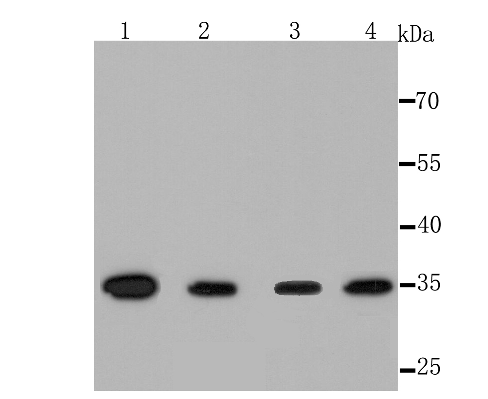
Fig1: Western blot analysis of Thymidylate Synthase on different cell lysate using anti-Thymidylate Synthase antibody at 1/1,000 dilution. Positive control: Lane1: Hela Lane2: Jurkat Lane3: Raji Lane4: Mouse testes Lane5: Mouse brain Lane6: Mouse spleenApplication
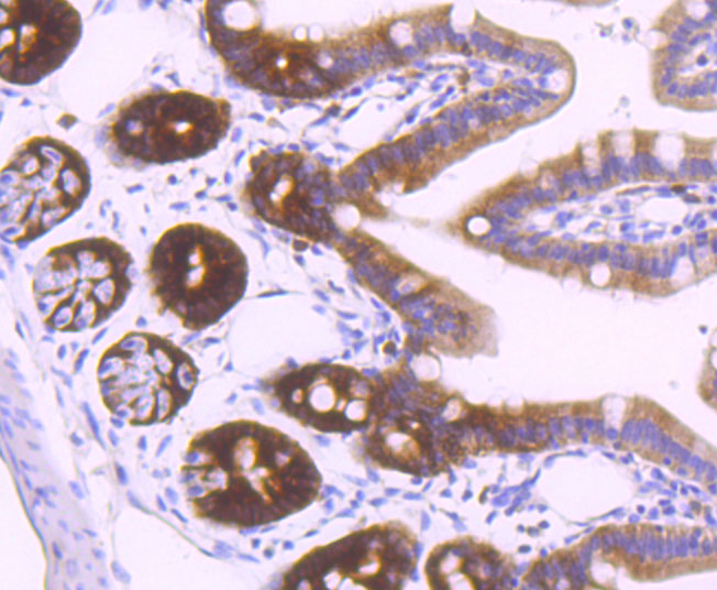
Fig2: Immunohistochemical analysis of paraffin-embedded mouse colon tissue using anti-Thymidylate Synthase antibody. Counter stained with hematoxylin.Application
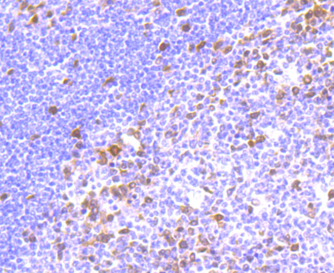
Fig3: Immunohistochemical analysis of paraffin-embedded human tonsil tissue using anti-Thymidylate Synthase antibody. Counter stained with hematoxylin.Application
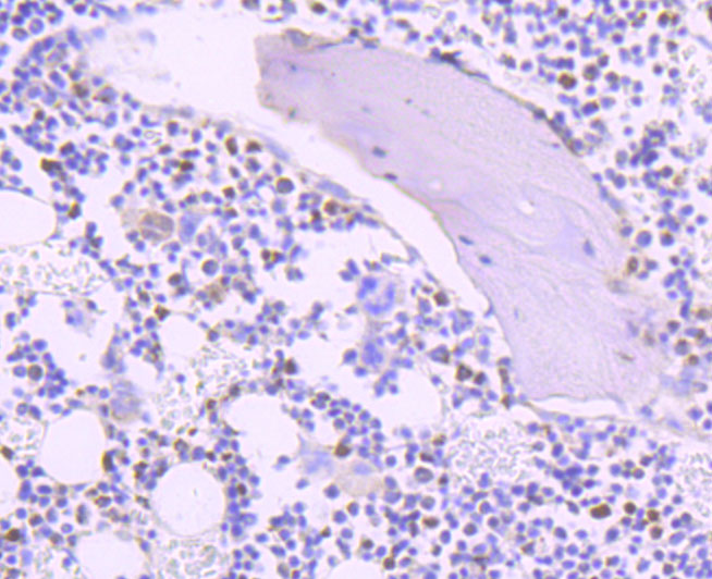
Fig4: Immunohistochemical analysis of paraffin-embedded rat bone tissue using anti-Thymidylate Synthase antibody. Counter stained with hematoxylin.Application
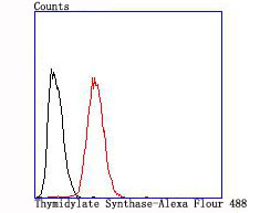
Fig5: Flow cytometric analysis of Hela cells with Thymidylate Synthase antibody at 1/100 dilution (red) compared with an unlabelled control (cells without incubation with primary antibody; black).| Product Name | Thymidylate Synthase [JM33-20] |
|---|---|
| Antibody Type | Primary Antibodies |
| Antigen Alias | d TMP synthase antibody EC 2.1.1.45 antibody HsT422 antibody MGC88736 antibody OTTHUMP00000162195 antibody Thymidylate synthase antibody Thymidylate synthetase antibody TMS antibody TS antibody TSase antibody Tyms antibody TYMS protein antibody Tyms thymidylate synthetase antibody TYSY_HUMAN antibody |
| Product description | Thymidylate synthase (TS), also designated TYMS, TMS, TSase and HsT422, uses 5,10-methylenetetrahydrofolate (methylene-THF) as a cofactor in the synthesis of 2'-deoxythymidine-5'-monophosphate (dTMP), an essential precursor for DNA biosynthesis. TS is an RNA-binding protein that can interact with its own mRNA. The TS/mRNA ribonucleoprotein complex can also associate with a number of other cellular mRNAs, including those corresponding to the p53 tumor suppressor gene and the Myc family of transcription factors. Inhibition of DNA replication and cell death resulting from thymidine depletion occurs when TS enzyme activity is inhibited with substrate or cofactor analogs, making the TS enzyme an important target for chemotherapy. Cancer cells are sensitive to thymidine depletion, as they multiply rapidly. |
| Immunogen | recombinant protein |
| Clonality | Monoclonal |
|---|---|
| Isotype | IgG |
| Host Species | Recombinant rabbit |
| Tested Applications | WBIHCIPFC |
| WB:1:500-1:2,000 IHC:1:50-1:200 FC:1:50-1:100 |
|
| Species Reactivity | HumanMouseRat |
| Concentration | 1mg/ml |
| Alternative Names | d TMP synthase antibody EC 2.1.1.45 antibody HsT422 antibody MGC88736 antibody OTTHUMP00000162195 antibody Thymidylate synthase antibody Thymidylate synthetase antibody TMS antibody TS antibody TSase antibody Tyms antibody TYMS protein antibody Tyms thymidylate synthetase antibody TYSY_HUMAN antibody |
|---|---|
| Molecular Weight(MW) | 36 kDa |
| Cellular Localization | Nucleus. Cytoplasm. Mitochondrion. |
Application
Fig1: Western blot analysis of Thymidylate Synthase on different cell lysate using anti-Thymidylate Synthase antibody at 1/1,000 dilution. Positive control: Lane1: Hela Lane2: Jurkat Lane3: Raji Lane4: Mouse testes Lane5: Mouse brain Lane6: Mouse spleen
Application
Fig2: Immunohistochemical analysis of paraffin-embedded mouse colon tissue using anti-Thymidylate Synthase antibody. Counter stained with hematoxylin.
Application
Fig3: Immunohistochemical analysis of paraffin-embedded human tonsil tissue using anti-Thymidylate Synthase antibody. Counter stained with hematoxylin.
Application
Fig4: Immunohistochemical analysis of paraffin-embedded rat bone tissue using anti-Thymidylate Synthase antibody. Counter stained with hematoxylin.
Application
Fig5: Flow cytometric analysis of Hela cells with Thymidylate Synthase antibody at 1/100 dilution (red) compared with an unlabelled control (cells without incubation with primary antibody; black).| Positive Control | Hela, Jurkat, Raji, mouse thymus, rat bone tissue, human tonsil tissue, mouse colon tissue. |
|---|---|
| Application Notes | WB:1:500-1:2,000 IHC:1:50-1:200 FC:1:50-1:100 |
| Form | Liquid |
|---|---|
| Storage Instructions | Store at +4℃ after thawing. Aliquot store at -20℃ or -80℃. Avoid repeated freeze / thaw cycles. |
| Storage Buffer | 1*TBS (pH7.4), 1%BSA, 40%Glycerol. Preservative: 0.05% Sodium Azide. |


