Application
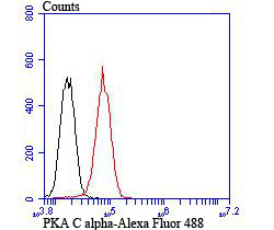
Fig6: Flow cytometric analysis of PC-3M cells with PKA C-alpha antibody at 1/100 dilution (red) compared with an unlabelled control (cells without incubation with primary antibody; black).Application
Fig6: Flow cytometric analysis of PC-3M cells with PKA C-alpha antibody at 1/100 dilution (red) compared with an unlabelled control (cells without incubation with primary antibody; black).Application
Fig6: Flow cytometric analysis of PC-3M cells with PKA C-alpha antibody at 1/100 dilution (red) compared with an unlabelled control (cells without incubation with primary antibody; black).Application
Fig6: Flow cytometric analysis of PC-3M cells with PKA C-alpha antibody at 1/100 dilution (red) compared with an unlabelled control (cells without incubation with primary antibody; black).Application
Fig6: Flow cytometric analysis of PC-3M cells with PKA C-alpha antibody at 1/100 dilution (red) compared with an unlabelled control (cells without incubation with primary antibody; black).Application
Fig6: Flow cytometric analysis of PC-3M cells with PKA C-alpha antibody at 1/100 dilution (red) compared with an unlabelled control (cells without incubation with primary antibody; black).| Product Name | PKA C alpha |
|---|---|
| Antibody Type | Primary Antibodies |
| Antigen Alias | cAMP dependent protein kinase beta catalytic subunit antibody cAMP dependent protein kinase alpha catalytic subunit antibody cAMP dependent protein kinase catalytic subunit alpha antibody cAMP dependent protein kinase catalytic subunit beta antibody PKA C alpha antibody PKA C beta antibody PKACA antibody PKACB antibody PRKACA antibody PRKACB antibody Protein kinase cAMP dependent catalytic alpha antibody Protein kinase cAMP dependent catalytic beta antibody |
| Product description | The second messenger cyclic AMP (cAMP) mediates diverse cellular responses to external signals such as proliferation, ion transport, regulation of metabolism and gene transcription by activation of the cAMP-dependent protein kinase (cAPK or PKA). Activation of PKA occurs when cAMP binds to the two regulatory subunits of the tetrameric PKA holoenzyme resulting in release of active catalytic subunits. Three catalytic (C) subunits have been identified, designated Cα, Cβ and Cγ, that each represent specific gene products. Cα and Cβ are closely related (93% amino acid sequence similarity), whereas Cγ displays 83% and 79% similarity to Cα and Cβ, respectively. Activation of transcription upon elevation of cAMP levels results from translocation of PKA to the nucleus where it phosphorylates the transcription factor cAMP response element binding protein (CREB) on serine 133 which in turn leads to TFIIB binding to TATA-box-binding protein TBP1, thus linking phospho-CREB to the pol II transcription initiation complex. |
| Immunogen | Recombinant protein. |
| Clonality | Polyclonal |
|---|---|
| Isotype | IgG |
| Host Species | Rabbit |
| Tested Applications | ICCIHCFC |
| ICC:1:50-1:200 IHC:1:50-1:200 FC:1:50-1:100 |
|
| Species Reactivity | HumanMouseRat |
| Concentration | 1mg/ml |
| Alternative Names | cAMP dependent protein kinase beta catalytic subunit antibody cAMP dependent protein kinase alpha catalytic subunit antibody cAMP dependent protein kinase catalytic subunit alpha antibody cAMP dependent protein kinase catalytic subunit beta antibody PKA C alpha antibody PKA C beta antibody PKACA antibody PKACB antibody PRKACA antibody PRKACB antibody Protein kinase cAMP dependent catalytic alpha antibody Protein kinase cAMP dependent catalytic beta antibody |
|---|---|
| Molecular Weight(MW) | 40 kDa |
| Cellular Localization | Mitochondrion. Nucleus.Plasma membrane. |
Application
Fig6: Flow cytometric analysis of PC-3M cells with PKA C-alpha antibody at 1/100 dilution (red) compared with an unlabelled control (cells without incubation with primary antibody; black).
Application
Fig6: Flow cytometric analysis of PC-3M cells with PKA C-alpha antibody at 1/100 dilution (red) compared with an unlabelled control (cells without incubation with primary antibody; black).
Application
Fig6: Flow cytometric analysis of PC-3M cells with PKA C-alpha antibody at 1/100 dilution (red) compared with an unlabelled control (cells without incubation with primary antibody; black).
Application
Fig6: Flow cytometric analysis of PC-3M cells with PKA C-alpha antibody at 1/100 dilution (red) compared with an unlabelled control (cells without incubation with primary antibody; black).
Application
Fig6: Flow cytometric analysis of PC-3M cells with PKA C-alpha antibody at 1/100 dilution (red) compared with an unlabelled control (cells without incubation with primary antibody; black).
Application
Fig6: Flow cytometric analysis of PC-3M cells with PKA C-alpha antibody at 1/100 dilution (red) compared with an unlabelled control (cells without incubation with primary antibody; black).| Positive Control | A431, PC-3M, rat brain tissue, human kidney tissue, mouse testis tissue. |
|---|---|
| Application Notes | ICC:1:50-1:200 IHC:1:50-1:200 FC:1:50-1:100 |
| Form | Liquid |
|---|---|
| Storage Instructions | Store at +4℃ after thawing. Aliquot store at -20℃ or -80℃. Avoid repeated freeze / thaw cycles. |
| Storage Buffer | 1*TBS (pH7.4), 0.5%BSA, 50%Glycerol. Preservative: 0.05% Sodium Azide. |


