Application
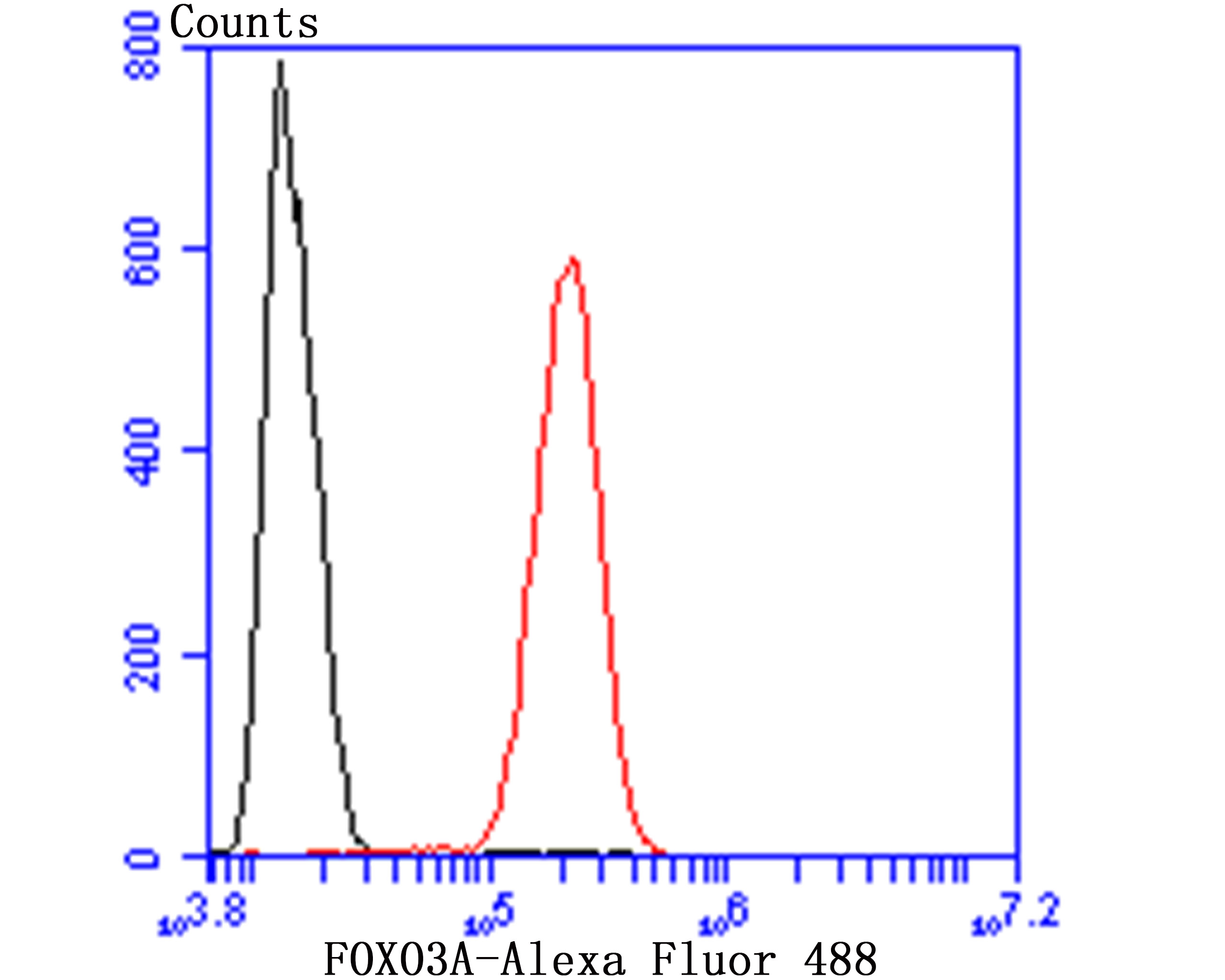
Fig8: Flow cytometric analysis of SHSY5Y cells with FOXO3A antibody at 1/50 dilution (red) compared with an unlabelled control (cells without incubation with primary antibody; black). Alexa Fluor 488-conjugated Goat anti rabbit IgG was used as the secondary antibody.Application
Fig8: Flow cytometric analysis of SHSY5Y cells with FOXO3A antibody at 1/50 dilution (red) compared with an unlabelled control (cells without incubation with primary antibody; black). Alexa Fluor 488-conjugated Goat anti rabbit IgG was used as the secondary antibody.Application
Fig8: Flow cytometric analysis of SHSY5Y cells with FOXO3A antibody at 1/50 dilution (red) compared with an unlabelled control (cells without incubation with primary antibody; black). Alexa Fluor 488-conjugated Goat anti rabbit IgG was used as the secondary antibody.Application
Fig8: Flow cytometric analysis of SHSY5Y cells with FOXO3A antibody at 1/50 dilution (red) compared with an unlabelled control (cells without incubation with primary antibody; black). Alexa Fluor 488-conjugated Goat anti rabbit IgG was used as the secondary antibody.Application
Fig8: Flow cytometric analysis of SHSY5Y cells with FOXO3A antibody at 1/50 dilution (red) compared with an unlabelled control (cells without incubation with primary antibody; black). Alexa Fluor 488-conjugated Goat anti rabbit IgG was used as the secondary antibody.Application
Fig8: Flow cytometric analysis of SHSY5Y cells with FOXO3A antibody at 1/50 dilution (red) compared with an unlabelled control (cells without incubation with primary antibody; black). Alexa Fluor 488-conjugated Goat anti rabbit IgG was used as the secondary antibody.Application
Fig8: Flow cytometric analysis of SHSY5Y cells with FOXO3A antibody at 1/50 dilution (red) compared with an unlabelled control (cells without incubation with primary antibody; black). Alexa Fluor 488-conjugated Goat anti rabbit IgG was used as the secondary antibody.Application
Fig8: Flow cytometric analysis of SHSY5Y cells with FOXO3A antibody at 1/50 dilution (red) compared with an unlabelled control (cells without incubation with primary antibody; black). Alexa Fluor 488-conjugated Goat anti rabbit IgG was used as the secondary antibody.| Product Name | FOXO3A |
|---|---|
| Antibody Type | Primary Antibodies |
| Antigen Alias | AF6q21 antibody AF6q21 protein antibody DKFZp781A0677 antibody FKHR2 antibody FKHRL 1 antibody FKHRL1 antibody FKHRL1P2 antibody Forkhead (Drosophila) homolog (rhabdomyosarcoma) like 1 antibody Forkhead box O3 antibody Forkhead box O3A antibody Forkhead box protein O3 antibody Forkhead box protein O3A antibody Forkhead Drosophila homolog of in rhabdomyosarcoma like 1 antibody Forkhead homolog (rhabdomyosarcoma) like 1 antibody Forkhead in rhabdomyosarcoma like 1 antibody Forkhead in rhabdomyosarcoma-like 1 antibody FOX O3A antibody FOXO2 antibody foxo3 antibody FOXO3_HUMAN antibody FOXO3A antibody MGC12739 antibody MGC31925 antibody |
| Product description | Transcriptional activator which triggers apoptosis in the absence of survival factors, including neuronal cell death upon oxidative stress. Recognizes and binds to the DNA sequence 5'-[AG]TAAA[TC]A-3'. Participates in post-transcriptional regulation of MYC: following phosphorylation by MAPKAPK5, promotes induction of miR-34b and miR-34c expression, 2 post-transcriptional regulators of MYC that bind to the 3'UTR of MYC transcript and prevent its translation. |
| Immunogen | Peptide |
| Clonality | Polyclonal |
|---|---|
| Isotype | IgG |
| Host Species | Rabbit |
| Tested Applications | WBICCIHCFC |
| WB:1:500-1:1,000 ICC:1:50-1:200 IHC:1:50-1:200 FC:1:50-1:100 |
|
| Species Reactivity | HumanMouse |
| Concentration | 1mg/ml |
| Alternative Names | AF6q21 antibody AF6q21 protein antibody DKFZp781A0677 antibody FKHR2 antibody FKHRL 1 antibody FKHRL1 antibody FKHRL1P2 antibody Forkhead (Drosophila) homolog (rhabdomyosarcoma) like 1 antibody Forkhead box O3 antibody Forkhead box O3A antibody Forkhead box protein O3 antibody Forkhead box protein O3A antibody Forkhead Drosophila homolog of in rhabdomyosarcoma like 1 antibody Forkhead homolog (rhabdomyosarcoma) like 1 antibody Forkhead in rhabdomyosarcoma like 1 antibody Forkhead in rhabdomyosarcoma-like 1 antibody FOX O3A antibody FOXO2 antibody foxo3 antibody FOXO3_HUMAN antibody FOXO3A antibody MGC12739 antibody MGC31925 antibody |
|---|---|
| Molecular Weight(MW) | 71 kDa |
| Cellular Localization | Cytoplasm. Nucleus. |
Application
Fig8: Flow cytometric analysis of SHSY5Y cells with FOXO3A antibody at 1/50 dilution (red) compared with an unlabelled control (cells without incubation with primary antibody; black). Alexa Fluor 488-conjugated Goat anti rabbit IgG was used as the secondary antibody.
Application
Fig8: Flow cytometric analysis of SHSY5Y cells with FOXO3A antibody at 1/50 dilution (red) compared with an unlabelled control (cells without incubation with primary antibody; black). Alexa Fluor 488-conjugated Goat anti rabbit IgG was used as the secondary antibody.
Application
Fig8: Flow cytometric analysis of SHSY5Y cells with FOXO3A antibody at 1/50 dilution (red) compared with an unlabelled control (cells without incubation with primary antibody; black). Alexa Fluor 488-conjugated Goat anti rabbit IgG was used as the secondary antibody.
Application
Fig8: Flow cytometric analysis of SHSY5Y cells with FOXO3A antibody at 1/50 dilution (red) compared with an unlabelled control (cells without incubation with primary antibody; black). Alexa Fluor 488-conjugated Goat anti rabbit IgG was used as the secondary antibody.
Application
Fig8: Flow cytometric analysis of SHSY5Y cells with FOXO3A antibody at 1/50 dilution (red) compared with an unlabelled control (cells without incubation with primary antibody; black). Alexa Fluor 488-conjugated Goat anti rabbit IgG was used as the secondary antibody.
Application
Fig8: Flow cytometric analysis of SHSY5Y cells with FOXO3A antibody at 1/50 dilution (red) compared with an unlabelled control (cells without incubation with primary antibody; black). Alexa Fluor 488-conjugated Goat anti rabbit IgG was used as the secondary antibody.
Application
Fig8: Flow cytometric analysis of SHSY5Y cells with FOXO3A antibody at 1/50 dilution (red) compared with an unlabelled control (cells without incubation with primary antibody; black). Alexa Fluor 488-conjugated Goat anti rabbit IgG was used as the secondary antibody.
Application
Fig8: Flow cytometric analysis of SHSY5Y cells with FOXO3A antibody at 1/50 dilution (red) compared with an unlabelled control (cells without incubation with primary antibody; black). Alexa Fluor 488-conjugated Goat anti rabbit IgG was used as the secondary antibody.| Positive Control | SH-SY5Y, K562, A431, LOVO, human tonsil tissue, mouse testis tissue, mouse fallopian tissue, SHSY5Y. |
|---|---|
| Application Notes | WB:1:500-1:1,000 ICC:1:50-1:200 IHC:1:50-1:200 FC:1:50-1:100 |
| Form | Liquid |
|---|---|
| Storage Instructions | Store at +4℃ after thawing. Aliquot store at -20℃ or -80℃. Avoid repeated freeze / thaw cycles. |
| Storage Buffer | 1*TBS (pH7.4), 0.5%BSA, 50%Glycerol. Preservative: 0.05% Sodium Azide. |


