Application
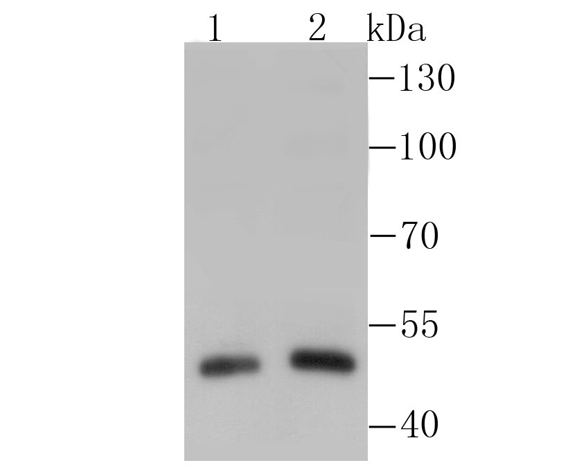
Fig1: Western blot analysis of RAR alpha on different lysates using anti-RAR alpha antibody at 1/500 dilution. Positive control: Lane1: Hela Lane2: Human lung tissueApplication
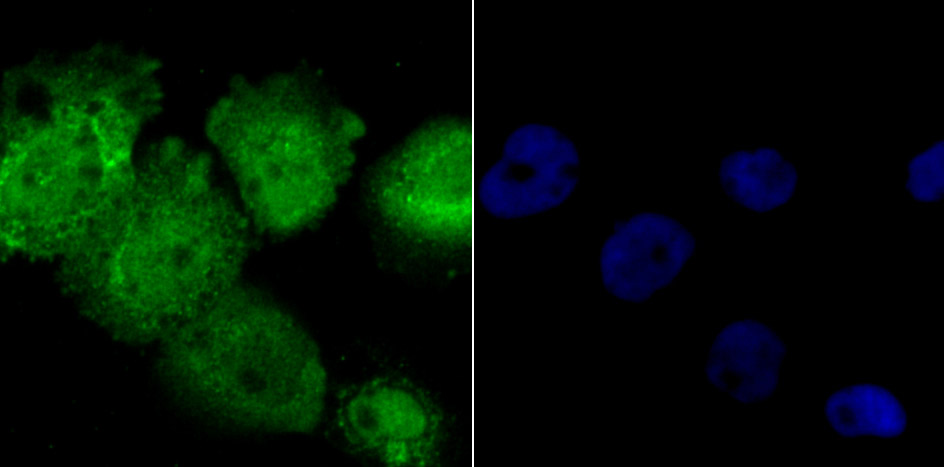
Fig2: ICC staining RAR alpha in A431 cells (green). The nuclear counter stain is DAPI (blue). Cells were fixed in paraformaldehyde, permeabilised with 0.25% Triton X100/PBS.Application
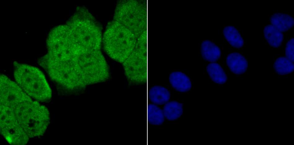
Fig3: ICC staining RAR alpha in MCF-7 cells (green). The nuclear counter stain is DAPI (blue). Cells were fixed in paraformaldehyde, permeabilised with 0.25% Triton X100/PBS.Application
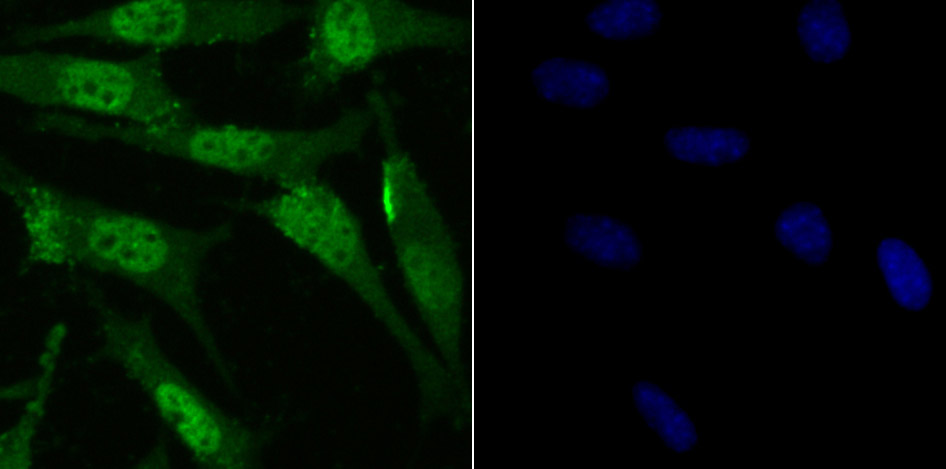
Fig4: ICC staining RAR alpha in SH-SY5Y cells (green). The nuclear counter stain is DAPI (blue). Cells were fixed in paraformaldehyde, permeabilised with 0.25% Triton X100/PBS.Application
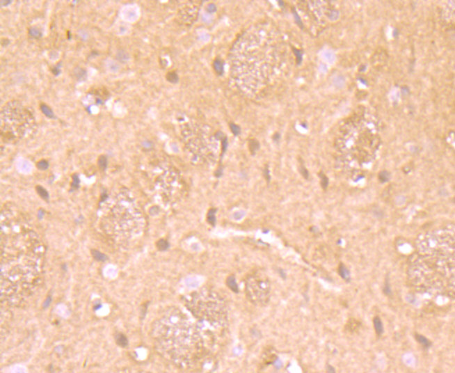
Fig5: Immunohistochemical analysis of paraffin-embedded rat brain tissue using anti-RAR alpha antibody. Counter stained with hematoxylin.Application
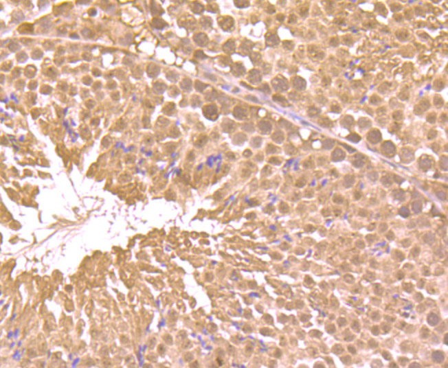
Fig6: Immunohistochemical analysis of paraffin-embedded mouse testis tissue using anti-RAR alpha antibody. Counter stained with hematoxylin.Application
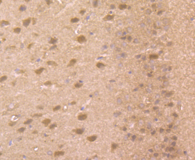
Fig7: Immunohistochemical analysis of paraffin-embedded mouse brian tissue using anti-RAR alpha antibody. Counter stained with hematoxylin.Application
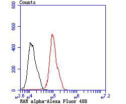
Fig8: Flow cytometric analysis of MCF-7 cells with RAR alpha antibody at 1/100 dilution (red) compared with an unlabelled control (cells without incubation with primary antibody; black). Alexa Fluor 488-conjugated Goat anti rabbit IgG was used as the secondary antibody.| Product Name | RAR alpha |
|---|---|
| Antibody Type | Primary Antibodies |
| Antigen Alias | NR1B1 antibody Nuclear mitotic apparatus protein retinoic acid receptor alpha fusion protein antibody Nuclear receptor subfamily 1 group B member 1 antibody Nucleophosmin retinoic acid receptor alpha fusion protein NPM RAR long form antibody RAR alpha antibody RAR antibody RAR-alpha antibody rara antibody RARA_HUMAN antibody RARalpha antibody RARalpha1 antibody Retinoic acid nuclear receptor alpha variant 1 antibody Retinoic acid nuclear receptor alpha variant 2 antibody Retinoic acid receptor alpha antibody Retinoic acid receptor alpha polypeptide antibody |
| Product description | A (retinol) that are important signaling molecules during vertebrate development and tissue differentiation. RAs activate the retinoic acid receptor (RAR) and retinoid X receptor (RXR) nuclear transcription factor families. Most retinoid forms activate RAR family members, whereas RXR family members are activated by 9-cis-RA only. RAR family members, which include RARα, RARβ and RARγ, have a high affinity for all transretinoic acids and belong to the same class of nuclear transcription factors as thyroid hormone receptors, vitamin D3 receptor and ecdysone receptor. RAR isoforms are expressed in distinct patterns throughout development and in the mature organism. The human RARα gene maps to chromosome 17 and is implicated in the chromosomal translocation associated with acute promyelocytic leukemia (APL-M3). Specifically, the RARα gene is fused with the promyelocytic leukemia (PML) gene, which encodes the fusion protein PML/RARα. The PML/RARα fusion protein inhibits PML-dependent apoptotic pathways and halts myeloid differentiation at the promyelocytic stage. |
| Immunogen | Recombinant protein |
| Clonality | Polyclonal |
|---|---|
| Isotype | IgG |
| Host Species | Rabbit |
| Tested Applications | WBICCIHCFC |
| WB:1:500 ICC:1:500-1:2,000 IHC:1:50-1:200 FC:1:50-1:100 |
|
| Species Reactivity | HumanMouseRat |
| Concentration | 1mg/ml |
| Alternative Names | NR1B1 antibody Nuclear mitotic apparatus protein retinoic acid receptor alpha fusion protein antibody Nuclear receptor subfamily 1 group B member 1 antibody Nucleophosmin retinoic acid receptor alpha fusion protein NPM RAR long form antibody RAR alpha antibody RAR antibody RAR-alpha antibody rara antibody RARA_HUMAN antibody RARalpha antibody RARalpha1 antibody Retinoic acid nuclear receptor alpha variant 1 antibody Retinoic acid nuclear receptor alpha variant 2 antibody Retinoic acid receptor alpha antibody Retinoic acid receptor alpha polypeptide antibody |
|---|---|
| Molecular Weight(MW) | 51 kDa |
| Cellular Localization | Nucleus. Cytoplasm. |
Application
Fig1: Western blot analysis of RAR alpha on different lysates using anti-RAR alpha antibody at 1/500 dilution. Positive control: Lane1: Hela Lane2: Human lung tissue
Application
Fig2: ICC staining RAR alpha in A431 cells (green). The nuclear counter stain is DAPI (blue). Cells were fixed in paraformaldehyde, permeabilised with 0.25% Triton X100/PBS.
Application
Fig3: ICC staining RAR alpha in MCF-7 cells (green). The nuclear counter stain is DAPI (blue). Cells were fixed in paraformaldehyde, permeabilised with 0.25% Triton X100/PBS.
Application
Fig4: ICC staining RAR alpha in SH-SY5Y cells (green). The nuclear counter stain is DAPI (blue). Cells were fixed in paraformaldehyde, permeabilised with 0.25% Triton X100/PBS.
Application
Fig5: Immunohistochemical analysis of paraffin-embedded rat brain tissue using anti-RAR alpha antibody. Counter stained with hematoxylin.
Application
Fig6: Immunohistochemical analysis of paraffin-embedded mouse testis tissue using anti-RAR alpha antibody. Counter stained with hematoxylin.
Application
Fig7: Immunohistochemical analysis of paraffin-embedded mouse brian tissue using anti-RAR alpha antibody. Counter stained with hematoxylin.
Application
Fig8: Flow cytometric analysis of MCF-7 cells with RAR alpha antibody at 1/100 dilution (red) compared with an unlabelled control (cells without incubation with primary antibody; black). Alexa Fluor 488-conjugated Goat anti rabbit IgG was used as the secondary antibody.| Positive Control | Hela, human lung tissue lysate, MCF-7, SH-SY5Y, rat brain tissue, mouse testis tissue, mouse brain tissue. |
|---|---|
| Application Notes | WB:1:500 ICC:1:500-1:2,000 IHC:1:50-1:200 FC:1:50-1:100 |
| Form | Liquid |
|---|---|
| Storage Instructions | Store at +4℃ after thawing. Aliquot store at -20℃ or -80℃. Avoid repeated freeze / thaw cycles. |
| Storage Buffer | 1*TBS (pH7.4), 0.5%BSA, 50%Glycerol. Preservative: 0.05% Sodium Azide. |


