Application
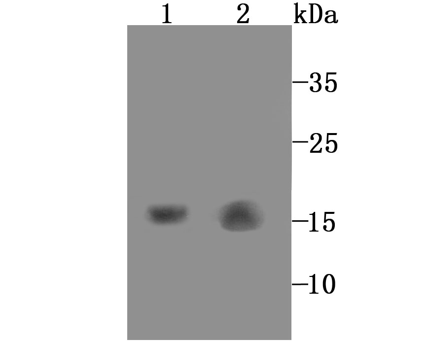
Fig1: Western blot analysis of GABARAP on mouse kidney (1) and rat liver (2) tissue lysate using anti-GABARAP antibody at 1/1,000 dilution.Application
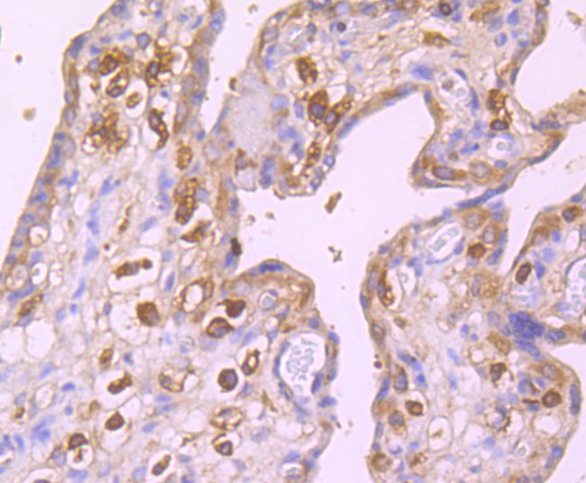
Fig2: Immunohistochemical analysis of paraffin-embedded human placenta tissue using anti-GABARAP antibody. Counter stained with hematoxylin.Application
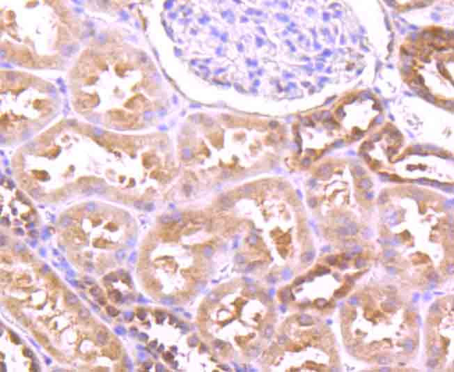
Fig3: Immunohistochemical analysis of paraffin-embedded human kidney tissue using anti-GABARAP antibody. Counter stained with hematoxylin.Application
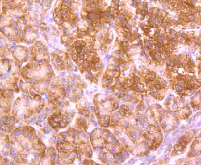
Fig4: Immunohistochemical analysis of paraffin-embedded human pancreas tissue using anti-GABARAP antibody. Counter stained with hematoxylin.Application
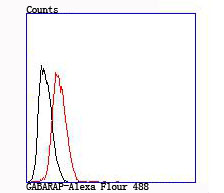
Fig5: Flow cytometric analysis of Hela cells with GABARAP antibody at 1/100 dilution (red) compared with an unlabelled control (cells without incubation with primary antibody; black).| Product Name | GABARAP [JM30-30] |
|---|---|
| Antibody Type | Primary Antibodies |
| Antigen Alias | ATG8A antibody FLC 3B antibody FLC3B antibody FLJ25768 antibody GABA type A receptor associated protein antibody GABA(A) receptor associated protein antibody GABA(A) receptor-associated protein antibody GABARAP a antibody GABARAP antibody Gamma aminobutyric acid receptor associated protein antibody Gamma-aminobutyric acid receptor-associated protein antibody GBRAP_HUMAN antibody MGC120154 antibody MGC120155 antibody MM 46 antibody MM46 antibody |
| Product description | In the central nervous system GABA functions as the main inhibitory transmitter by increasing a Cl-conductance that inhibits neuronal firing. GABA has been shown to activate both ionotropic (GABAA) and metabotropic (GABAB) receptors as well as a third class of receptors called GABAC. In addition to GABA receptors, several proteins have been identified as regulators of GABA function, including GAD65, GAD67, GABA transporters and GABARAP (GABAA receptor-associated protein). GABARAP associates with GABAA Rg2 to link GABAA receptors to the cytoskeleton. The GABARAP protein sequence is similar to light chain-3 of microtubule-associated proteins (MAPs) suggesting that it may be a type of MAP or a component of a MAP complex. |
| Immunogen | Recombinant protein |
| Clonality | Monoclonal |
|---|---|
| Isotype | IgG |
| Host Species | Recombinant rabbit |
| Tested Applications | WBIHCFC |
| WB:1:500 IHC:1:50-1:200 FC:1:50-1:100 |
|
| Species Reactivity | HumanMouseRat |
| Concentration | 1mg/ml |
| Alternative Names | ATG8A antibody FLC 3B antibody FLC3B antibody FLJ25768 antibody GABA type A receptor associated protein antibody GABA(A) receptor associated protein antibody GABA(A) receptor-associated protein antibody GABARAP a antibody GABARAP antibody Gamma aminobutyric acid receptor associated protein antibody Gamma-aminobutyric acid receptor-associated protein antibody GBRAP_HUMAN antibody MGC120154 antibody MGC120155 antibody MM 46 antibody MM46 antibody |
|---|---|
| Molecular Weight(MW) | 14 kDa |
| Cellular Localization | Endomembrane system. Cytoplasm, cytoskeleton. |
Application
Fig1: Western blot analysis of GABARAP on mouse kidney (1) and rat liver (2) tissue lysate using anti-GABARAP antibody at 1/1,000 dilution.
Application
Fig2: Immunohistochemical analysis of paraffin-embedded human placenta tissue using anti-GABARAP antibody. Counter stained with hematoxylin.
Application
Fig3: Immunohistochemical analysis of paraffin-embedded human kidney tissue using anti-GABARAP antibody. Counter stained with hematoxylin.
Application
Fig4: Immunohistochemical analysis of paraffin-embedded human pancreas tissue using anti-GABARAP antibody. Counter stained with hematoxylin.
Application
Fig5: Flow cytometric analysis of Hela cells with GABARAP antibody at 1/100 dilution (red) compared with an unlabelled control (cells without incubation with primary antibody; black).| Positive Control | Mouse kidney tissue lysate, rat liver tissue lysate, Hela, human placenta tissue, human kidney tissue, human pancreas tissue. |
|---|---|
| Application Notes | WB:1:500 IHC:1:50-1:200 FC:1:50-1:100 |
| Form | Liquid |
|---|---|
| Storage Instructions | Store at +4℃ after thawing. Aliquot store at -20℃ or -80℃. Avoid repeated freeze / thaw cycles. |
| Storage Buffer | 1*TBS (pH7.4), 1%BSA, 40%Glycerol. Preservative: 0.05% Sodium Azide. |


