Application
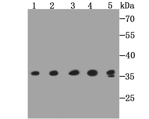
Fig1: Western blot analysis of GAPDH on different lysates using anti-GAPDH antibody at 1/2,000 dilution. Positive control: Lane1: PC-12 Lane2: L929 Lane3: NIH/3T3 Lane4: F9 Lane5: Mouse liver tissueApplication
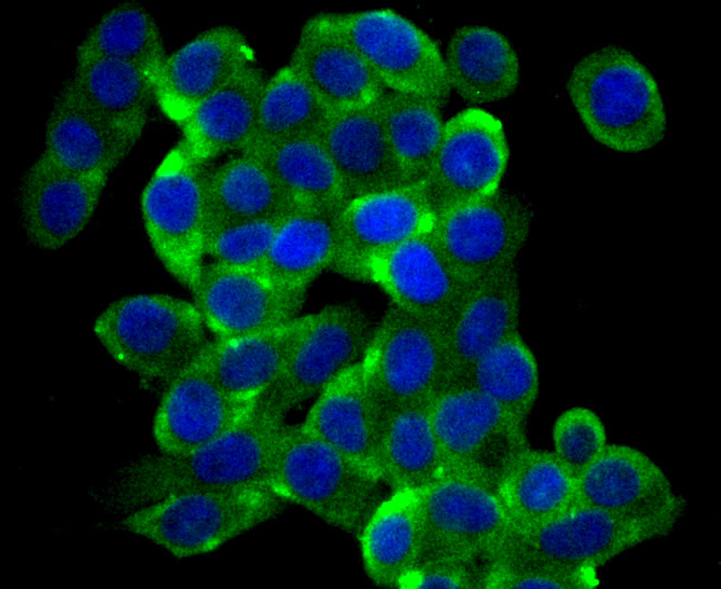
Fig2: ICC staining GAPDH in LOVO cells (green). The nuclear counter stain is DAPI (blue). Cells were fixed in paraformaldehyde, permeabilised with 0.25% Triton X100/PBS.Application
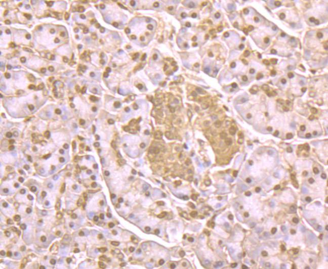
Fig3: Immunohistochemical analysis of paraffin-embedded human pancreas tissue using anti-GAPDH antibody. Counter stained with hematoxylin.Application
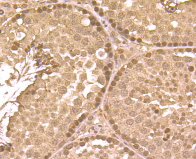
Fig4: Immunohistochemical analysis of paraffin-embedded mouse testis tissue using anti-GAPDH antibody. Counter stained with hematoxylin.Application
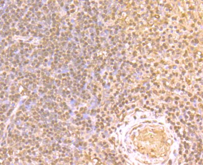
Fig5: Immunohistochemical analysis of paraffin-embedded human tonsil tissue using anti-GAPDH antibody. Counter stained with hematoxylin.Application
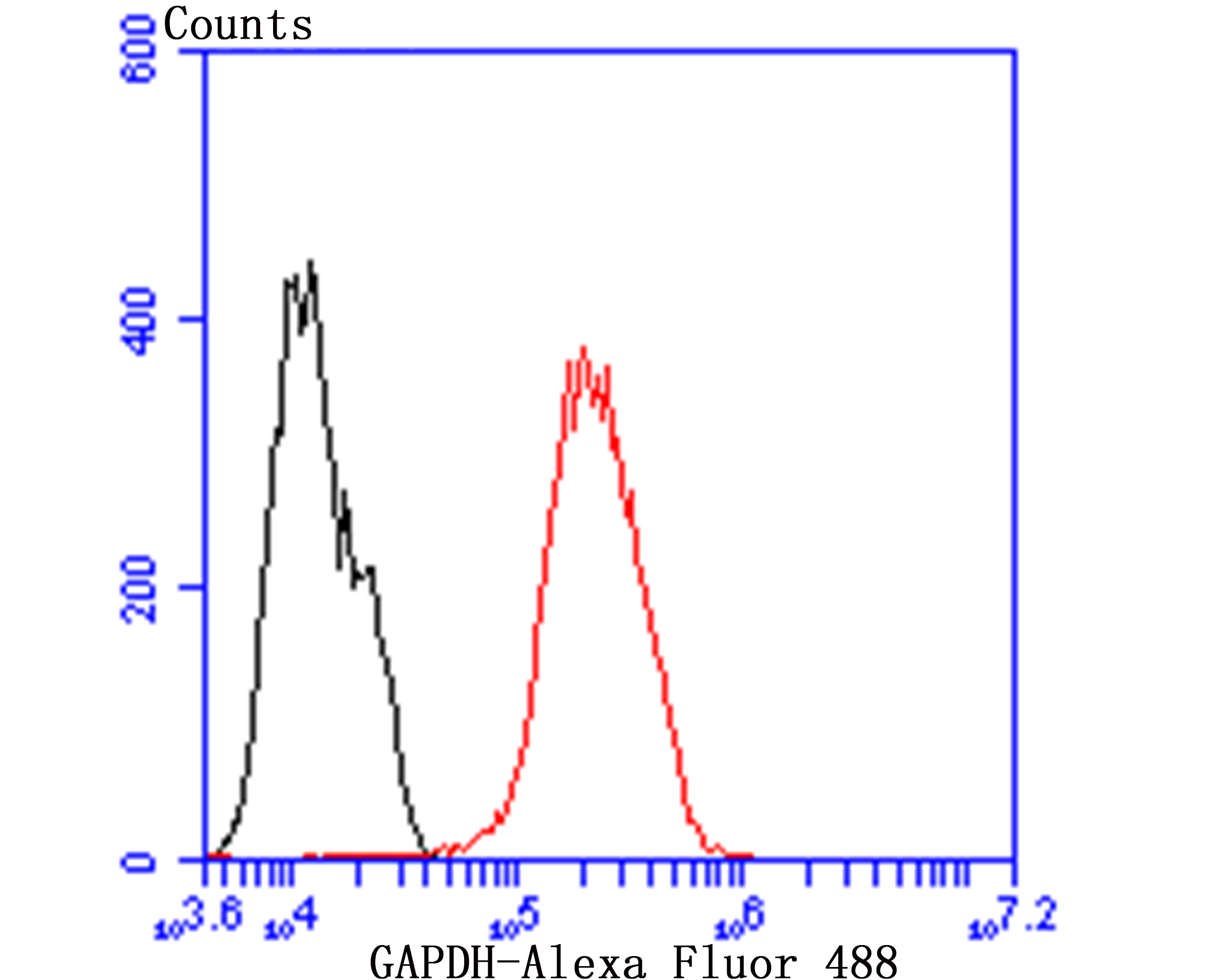
Fig6: Flow cytometric analysis of MCF-7 cells with GAPDH antibody at 1/50 dilution (red) compared with an unlabelled control (cells without incubation with primary antibody; black). Alexa Fluor 488-conjugated goat anti rabbit IgG was used as the secondary antibody.| Product Name | GAPDH |
|---|---|
| Antibody Type | Primary Antibodies |
| Antigen Alias | 38 kDa BFA-dependent ADP-ribosylation substrate antibody aging associated gene 9 protein antibody Aging-associated gene 9 protein antibody BARS-38 antibody cb609 antibody EC 1.2.1.12 antibody Epididymis secretory sperm binding protein Li 162eP antibody G3P_HUMAN antibody G3PD antibody G3PDH antibody GAPD antibody GAPDH antibody Glyceraldehyde 3 phosphate dehydrogenase antibody Glyceraldehyde-3-phosphate dehydrogenase antibody HEL-S-162eP antibody KNC-NDS6 antibody MGC102544 antibody MGC102546 antibody MGC103190 antibody MGC103191 antibody MGC105239 antibody MGC127711 antibody MGC88685 antibody OCAS, p38 component antibody OCT1 coactivator in S phase, 38-KD component antibody peptidyl cysteine S nitrosylase GAPDH antibody Peptidyl-cysteine S-nitrosylase GAPDH antibody wu:fb33a10 antibody |
| Product description | Has both glyceraldehyde-3-phosphate dehydrogenase and nitrosylase activities, thereby playing a role in glycolysis and nuclear functions, respectively. Participates in nuclear events including transcription, RNA transport, DNA replication and apoptosis. Nuclear functions are probably due to the nitrosylase activity that mediates cysteine S-nitrosylation of nuclear target proteins such as SIRT1, HDAC2 and PRKDC. Modulates the organization and assembly of the cytoskeleton. Facilitates the CHP1-dependent microtubule and membrane associations through its ability to stimulate the binding of CHP1 to microtubules (By similarity). Glyceraldehyde-3-phosphate dehydrogenase is a key enzyme in glycolysis that catalyzes the first step of the pathway by converting D-glyceraldehyde 3-phosphate (G3P) into 3-phospho-D-glyceroyl phosphate. Component of the GAIT (gamma interferon-activated inhibitor of translation) complex which mediates interferon-gamma-induced transcript-selective translation inhibition in inflammation processes. |
| Immunogen | Peptide |
| Clonality | Polyclonal |
|---|---|
| Isotype | IgG |
| Host Species | Rabbit |
| Tested Applications | WBICCIHCFC |
| WB:1:2,000-5,000 ICC:1:50-1:200 IHC:1:50-1:200 FC:1:50-1 100 |
|
| Species Reactivity | HumanMouseRat |
| Concentration | 1mg/ml |
| Alternative Names | 38 kDa BFA-dependent ADP-ribosylation substrate antibody
aging associated gene 9 protein antibody
Aging-associated gene 9 protein antibody
BARS-38 antibody
cb609 antibody
EC 1.2.1.12 antibody
Epididymis secretory sperm binding protein Li 162eP antibody
G3P_HUMAN antibody
G3PD antibody
G3PDH antibody
GAPD antibody
GAPDH antibody
Glyceraldehyde 3 phosphate dehydrogenase antibody
Glyceraldehyde-3-phosphate dehydrogenase antibody
HEL-S-162eP antibody
KNC-NDS6 antibody
MGC102544 antibody
MGC102546 antibody
MGC103190 antibody
MGC103191 antibody
MGC105239 antibody
MGC127711 antibody
MGC88685 antibody
OCAS p38 component antibody OCT1 coactivator in S phase 38-KD component antibody peptidyl cysteine S nitrosylase GAPDH antibody Peptidyl-cysteine S-nitrosylase GAPDH antibody wu:fb33a10 antibody |
|---|---|
| Molecular Weight(MW) | 36 kDa |
| Cellular Localization | Cytoskeleton. Cytosol. Nucleus. |
Application
Fig1: Western blot analysis of GAPDH on different lysates using anti-GAPDH antibody at 1/2,000 dilution. Positive control: Lane1: PC-12 Lane2: L929 Lane3: NIH/3T3 Lane4: F9 Lane5: Mouse liver tissue
Application
Fig2: ICC staining GAPDH in LOVO cells (green). The nuclear counter stain is DAPI (blue). Cells were fixed in paraformaldehyde, permeabilised with 0.25% Triton X100/PBS.
Application
Fig3: Immunohistochemical analysis of paraffin-embedded human pancreas tissue using anti-GAPDH antibody. Counter stained with hematoxylin.
Application
Fig4: Immunohistochemical analysis of paraffin-embedded mouse testis tissue using anti-GAPDH antibody. Counter stained with hematoxylin.
Application
Fig5: Immunohistochemical analysis of paraffin-embedded human tonsil tissue using anti-GAPDH antibody. Counter stained with hematoxylin.
Application
Fig6: Flow cytometric analysis of MCF-7 cells with GAPDH antibody at 1/50 dilution (red) compared with an unlabelled control (cells without incubation with primary antibody; black). Alexa Fluor 488-conjugated goat anti rabbit IgG was used as the secondary antibody.| Positive Control | PC-12, L929, NIH/3T3, F9, mouse liver tissue lysate, human tonsil tissue, human pancreas tissue, mouse testis tissue, MCF-7. |
|---|---|
| Application Notes | WB:1:2,000-5,000 ICC:1:50-1:200 IHC:1:50-1:200 FC:1:50-1 100 |
| Form | Liquid |
|---|---|
| Storage Instructions | Store at +4℃ after thawing. Aliquot store at -20℃ or -80℃. Avoid repeated freeze / thaw cycles. |
| Storage Buffer | 1*TBS (pH7.4), 0.5%BSA, 50%Glycerol. Preservative: 0.05% Sodium Azide. |


