Application
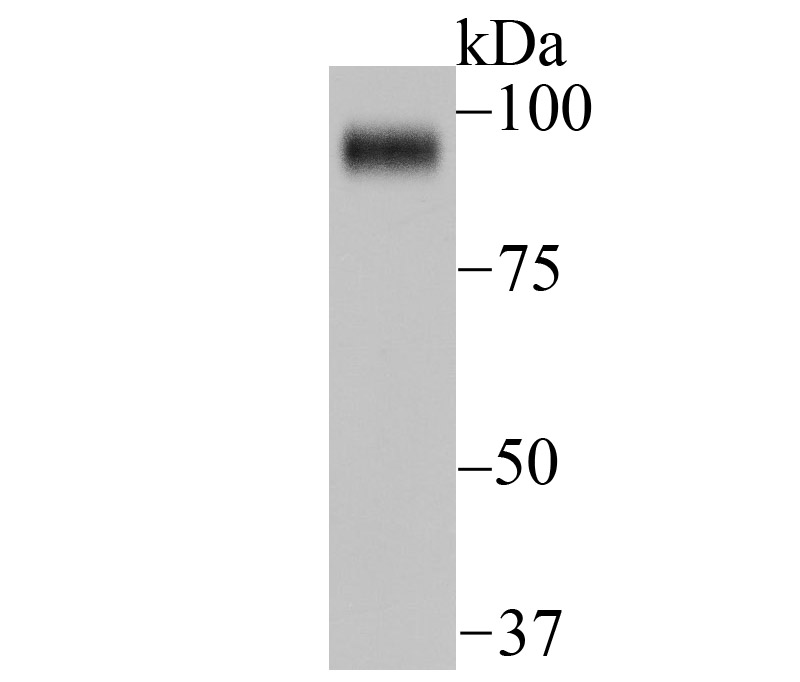
Fig1: Western blot analysis of FAP on Siha cell lysate using anti-FAP antibody at 1/1,000 dilution.Application
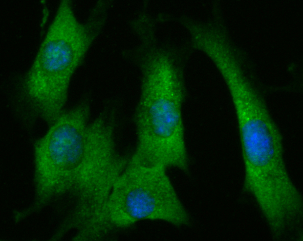
Fig2: ICC staining FAP in NIH-3T3 cells (green). The nuclear counter stain is DAPI (blue). Cells were fixed in paraformaldehyde, permeabilised with 0.25% Triton X100/PBS.Application
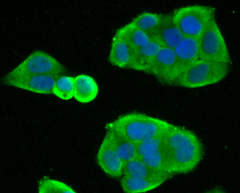
Fig3: ICC staining FAP in Hela cells (green). The nuclear counter stain is DAPI (blue). Cells were fixed in paraformaldehyde, permeabilised with 0.25% Triton X100/PBS.Application
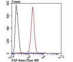
Fig4: Flow cytometric analysis of SH-SY5Y cells with FAP antibody at 1/100 dilution (red) compared with an unlabelled control (cells without incubation with primary antibody; black). Alexa Fluor 488-conjugated goat anti rabbit IgG was used as the secondary antibody.| Product Name | FAP |
|---|---|
| Antibody Type | Primary Antibodies |
| Antigen Alias | Adenomatous Polyposis Coli antibody Adenomatous polyposis coli protein antibody Apc antibody APC_HUMAN antibody CC1 antibody Deleted in polyposis 2.5 antibody DP2 antibody DP2.5 antibody DP3 antibody FAP antibody FPC antibody GS antibody Protein APC antibody |
| Product description | Cell surface glycoprotein serine protease that participates in extracellular matrix degradation and involved in many cellular processes including tissue remodeling, fibrosis, wound healing, inflammation and tumor growth. Plays a role in tissue remodeling during development and wound healing. Participates in the cell invasiveness towards the ECM in malignant melanoma cancers. Enhances tumor growth progression by increasing angiogenesis, collagen fiber degradation and apoptosis and by reducing antitumor response of the immune system. Promotes glioma cell invasion through the brain parenchyma by degrading the proteoglycan brevican. Acts as a tumor suppressor in melanocytic cells through regulation of cell proliferation and survival in a serine protease activity-independent manner. |
| Immunogen | Peptide |
| Clonality | Polyclonal |
|---|---|
| Isotype | IgG |
| Host Species | Rabbit |
| Tested Applications | WBICCFC |
| WB:1:500-1:1,000 ICC:1:50-1:200 FC:1:50-1:100 |
|
| Species Reactivity | HumanMouse |
| Concentration | 1mg/ml |
| Alternative Names | Adenomatous Polyposis Coli antibody Adenomatous polyposis coli protein antibody Apc antibody APC_HUMAN antibody CC1 antibody Deleted in polyposis 2.5 antibody DP2 antibody DP2.5 antibody DP3 antibody FAP antibody FPC antibody GS antibody Protein APC antibody |
|---|---|
| Molecular Weight(MW) | 95 kDa |
| Cellular Localization | Cell membrane. |
Application
Fig1: Western blot analysis of FAP on Siha cell lysate using anti-FAP antibody at 1/1,000 dilution.
Application
Fig2: ICC staining FAP in NIH-3T3 cells (green). The nuclear counter stain is DAPI (blue). Cells were fixed in paraformaldehyde, permeabilised with 0.25% Triton X100/PBS.
Application
Fig3: ICC staining FAP in Hela cells (green). The nuclear counter stain is DAPI (blue). Cells were fixed in paraformaldehyde, permeabilised with 0.25% Triton X100/PBS.
Application
Fig4: Flow cytometric analysis of SH-SY5Y cells with FAP antibody at 1/100 dilution (red) compared with an unlabelled control (cells without incubation with primary antibody; black). Alexa Fluor 488-conjugated goat anti rabbit IgG was used as the secondary antibody.| Positive Control | Hela, NIH-3T3, Siha, SH-SY5Y. |
|---|---|
| Application Notes | WB:1:500-1:1,000 ICC:1:50-1:200 FC:1:50-1:100 |
| Form | Liquid |
|---|---|
| Storage Instructions | Store at +4℃ after thawing. Aliquot store at -20℃ or -80℃. Avoid repeated freeze / thaw cycles. |
| Storage Buffer | 1*TBS (pH7.4), 0.5%BSA, 50%Glycerol. Preservative: 0.05% Sodium Azide. |


