Application
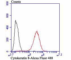
Fig9: Flow cytometric analysis of MCF-7 cells with Cytokeratin 8 antibody at 1/100 dilution (red) compared with an unlabelled control (cells without incubation with primary antibody; black).Application
Fig9: Flow cytometric analysis of MCF-7 cells with Cytokeratin 8 antibody at 1/100 dilution (red) compared with an unlabelled control (cells without incubation with primary antibody; black).Application
Fig9: Flow cytometric analysis of MCF-7 cells with Cytokeratin 8 antibody at 1/100 dilution (red) compared with an unlabelled control (cells without incubation with primary antibody; black).Application
Fig9: Flow cytometric analysis of MCF-7 cells with Cytokeratin 8 antibody at 1/100 dilution (red) compared with an unlabelled control (cells without incubation with primary antibody; black).Application
Fig9: Flow cytometric analysis of MCF-7 cells with Cytokeratin 8 antibody at 1/100 dilution (red) compared with an unlabelled control (cells without incubation with primary antibody; black).Application
Fig9: Flow cytometric analysis of MCF-7 cells with Cytokeratin 8 antibody at 1/100 dilution (red) compared with an unlabelled control (cells without incubation with primary antibody; black).Application
Fig9: Flow cytometric analysis of MCF-7 cells with Cytokeratin 8 antibody at 1/100 dilution (red) compared with an unlabelled control (cells without incubation with primary antibody; black).Application
Fig9: Flow cytometric analysis of MCF-7 cells with Cytokeratin 8 antibody at 1/100 dilution (red) compared with an unlabelled control (cells without incubation with primary antibody; black).Application
Fig9: Flow cytometric analysis of MCF-7 cells with Cytokeratin 8 antibody at 1/100 dilution (red) compared with an unlabelled control (cells without incubation with primary antibody; black).| Product Name | Cytokeratin 8 |
|---|---|
| Antibody Type | Primary Antibodies |
| Antigen Alias | CARD2 antibody CK 8 antibody CK-8 antibody CK8 antibody CYK8 antibody CYKER antibody Cytokeratin endo A antibody Cytokeratin-8 antibody DreK8 antibody EndoA antibody K2C8 antibody K2C8_HUMAN antibody K8 antibody Keratin 8 antibody Keratin type II cytoskeletal 8 antibody Keratin, type II cytoskeletal 8 antibody Keratin-8 antibody KO antibody Krt 2.8 antibody KRT8 antibody MGC118110 antibody MGC174782 antibody MGC53564 antibody MGC85764 antibody sb:cb186 antibody Type-II keratin Kb8 antibody |
| Product description | Cytokeratins comprise a diverse group of intermediate filament proteins (IFPs) that are expressed as pairs in both keratinized and non-keratinized epithelial tissue. Cytokeratins play a critical role in differentiation and tissue specialization and function to maintain the overall structural integrity of epithelial cells. They have been found to be useful markers of tissue differentiation, which is directly applicable to the characterization of malignant tumors. Cytokeratin 8 expression is seen in epithelium and epithelium-derived tumors. The Cytokeratin 8 and 18 pair are normally expressed in simple epithelia, but not in stratified epithelial cells. Research indicates that squamous cell carcinomas derived from stratified epithelia show abnormal expression of Cytokeratin 8 and 18, although it is not known whether these proteins contribute to the malignant phenotype of the cells. Expression of Cytokeratin 8 and 18 in oral squamous cell carcinomas is an independent prognostic marker that indicates a poor prognosis. Cytokeratin 8 expression correlates with malignancy in leukoplakia and carcinomas of the head and neck; it is expressed in all non-small-cell lung cancers. Cytokeratin 8 has been shown to possess extracellular epitopes on tumor cells, which may represent valuable targets for therapy. |
| Immunogen | Peptide |
| Clonality | Polyclonal |
|---|---|
| Isotype | IgG |
| Host Species | Rabbit |
| Tested Applications | WBICCIHCFC |
| WB:1:500 ICC:1:100-1:500 IHC:1:50-1:200 FC:1:50-1:100 |
|
| Species Reactivity | HumanMouseRat |
| Concentration | 1mg/ml |
| Alternative Names | CARD2 antibody
CK 8 antibody
CK-8 antibody
CK8 antibody
CYK8 antibody
CYKER antibody
Cytokeratin endo A antibody
Cytokeratin-8 antibody
DreK8 antibody
EndoA antibody
K2C8 antibody
K2C8_HUMAN antibody
K8 antibody
Keratin 8 antibody
Keratin type II cytoskeletal 8 antibody
Keratin type II cytoskeletal 8 antibody Keratin-8 antibody KO antibody Krt 2.8 antibody KRT8 antibody MGC118110 antibody MGC174782 antibody MGC53564 antibody MGC85764 antibody sb:cb186 antibody Type-II keratin Kb8 antibody |
|---|---|
| Molecular Weight(MW) | 53 kDa |
| Cellular Localization | Cytoplasm. Nucleus. |
Application
Fig9: Flow cytometric analysis of MCF-7 cells with Cytokeratin 8 antibody at 1/100 dilution (red) compared with an unlabelled control (cells without incubation with primary antibody; black).
Application
Fig9: Flow cytometric analysis of MCF-7 cells with Cytokeratin 8 antibody at 1/100 dilution (red) compared with an unlabelled control (cells without incubation with primary antibody; black).
Application
Fig9: Flow cytometric analysis of MCF-7 cells with Cytokeratin 8 antibody at 1/100 dilution (red) compared with an unlabelled control (cells without incubation with primary antibody; black).
Application
Fig9: Flow cytometric analysis of MCF-7 cells with Cytokeratin 8 antibody at 1/100 dilution (red) compared with an unlabelled control (cells without incubation with primary antibody; black).
Application
Fig9: Flow cytometric analysis of MCF-7 cells with Cytokeratin 8 antibody at 1/100 dilution (red) compared with an unlabelled control (cells without incubation with primary antibody; black).
Application
Fig9: Flow cytometric analysis of MCF-7 cells with Cytokeratin 8 antibody at 1/100 dilution (red) compared with an unlabelled control (cells without incubation with primary antibody; black).
Application
Fig9: Flow cytometric analysis of MCF-7 cells with Cytokeratin 8 antibody at 1/100 dilution (red) compared with an unlabelled control (cells without incubation with primary antibody; black).
Application
Fig9: Flow cytometric analysis of MCF-7 cells with Cytokeratin 8 antibody at 1/100 dilution (red) compared with an unlabelled control (cells without incubation with primary antibody; black).
Application
Fig9: Flow cytometric analysis of MCF-7 cells with Cytokeratin 8 antibody at 1/100 dilution (red) compared with an unlabelled control (cells without incubation with primary antibody; black).| Positive Control | Human small intestine tissue lysate, A549, LOVO, MCF-7, rat epididymis tissue, human colon cancer tissue, human breast tissue, mouse fallopian tubes tissue. |
|---|---|
| Application Notes | WB:1:500 ICC:1:100-1:500 IHC:1:50-1:200 FC:1:50-1:100 |
| Form | Liquid |
|---|---|
| Storage Instructions | Store at +4℃ after thawing. Aliquot store at -20℃ or -80℃. Avoid repeated freeze / thaw cycles. |
| Storage Buffer | 1*TBS (pH7.4), 0.5%BSA, 50%Glycerol. Preservative: 0.05% Sodium Azide. |


