Application
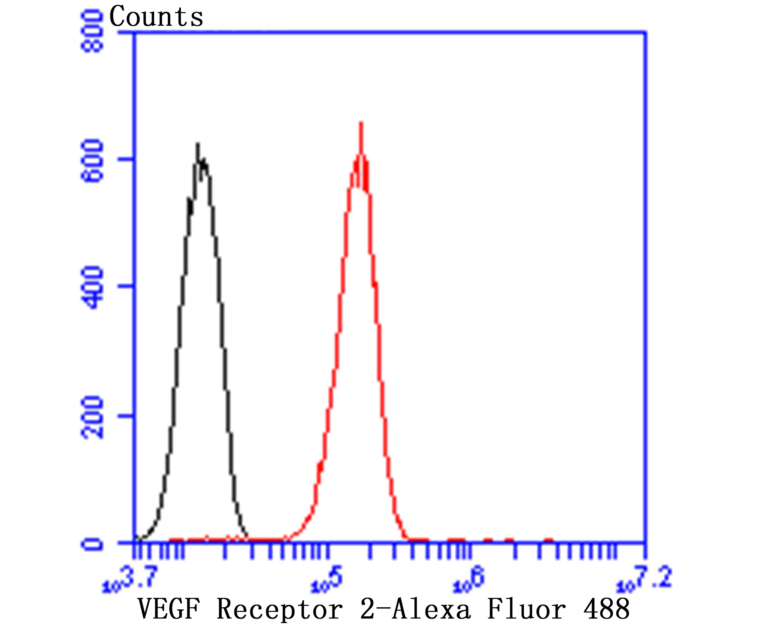
Fig8: Flow cytometric analysis of HUVEC cells with VEGF Receptor 2 antibody at 1/100 dilution (red) compared with an unlabelled control (cells without incubation with primary antibody; black). Alexa Fluor 488-conjugated goat anti rabbit IgG was used as the secondary antibody.Application
Fig8: Flow cytometric analysis of HUVEC cells with VEGF Receptor 2 antibody at 1/100 dilution (red) compared with an unlabelled control (cells without incubation with primary antibody; black). Alexa Fluor 488-conjugated goat anti rabbit IgG was used as the secondary antibody.Application
Fig8: Flow cytometric analysis of HUVEC cells with VEGF Receptor 2 antibody at 1/100 dilution (red) compared with an unlabelled control (cells without incubation with primary antibody; black). Alexa Fluor 488-conjugated goat anti rabbit IgG was used as the secondary antibody.Application
Fig8: Flow cytometric analysis of HUVEC cells with VEGF Receptor 2 antibody at 1/100 dilution (red) compared with an unlabelled control (cells without incubation with primary antibody; black). Alexa Fluor 488-conjugated goat anti rabbit IgG was used as the secondary antibody.Application
Fig8: Flow cytometric analysis of HUVEC cells with VEGF Receptor 2 antibody at 1/100 dilution (red) compared with an unlabelled control (cells without incubation with primary antibody; black). Alexa Fluor 488-conjugated goat anti rabbit IgG was used as the secondary antibody.Application
Fig8: Flow cytometric analysis of HUVEC cells with VEGF Receptor 2 antibody at 1/100 dilution (red) compared with an unlabelled control (cells without incubation with primary antibody; black). Alexa Fluor 488-conjugated goat anti rabbit IgG was used as the secondary antibody.Application
Fig8: Flow cytometric analysis of HUVEC cells with VEGF Receptor 2 antibody at 1/100 dilution (red) compared with an unlabelled control (cells without incubation with primary antibody; black). Alexa Fluor 488-conjugated goat anti rabbit IgG was used as the secondary antibody.Application
Fig8: Flow cytometric analysis of HUVEC cells with VEGF Receptor 2 antibody at 1/100 dilution (red) compared with an unlabelled control (cells without incubation with primary antibody; black). Alexa Fluor 488-conjugated goat anti rabbit IgG was used as the secondary antibody.| Product Name | VEGF Receptor 2 |
|---|---|
| Antibody Type | Primary Antibodies |
| Antigen Alias | CD309 antibody CD309 antigen antibody EC 2.7.10.1 antibody Fetal liver kinase 1 antibody FLK-1 antibody FLK1 antibody FLK1, mouse, homolog of antibody Kdr antibody Kinase insert domain receptor (a type III receptor tyrosine kinase) antibody Kinase insert domain receptor antibody KRD1 antibody Ly73 antibody Protein tyrosine kinase receptor FLK1 antibody Protein-tyrosine kinase receptor flk-1 antibody soluble VEGFR2 antibody Tyrosine kinase growth factor receptor antibody Vascular endothelial growth factor receptor 2 antibody VEGFR 2 antibody VEGFR antibody VEGFR-2 antibody VEGFR2 antibody VGFR2_HUMAN antibody |
| Product description | Three cell membrane receptor tyrosine kinases, Flt (also designated VEGF-R1) (1-3), Flk-1 (also designated VEGF-R2) (4-6) and Flt-4, putatively involved in the growth of endothelial cells, are characterized by the presence of seven immunoglobulin-like sequences in their extracellular domain. These receptors exhibit high degrees of sequence relatedness to each other as well as lesser degrees of relatedness to the class III receptors including CSF-1/Fms, PDGR, SLFR/Kit and Flt-3/Flk-2. Two members of this receptor class, Flt-1 and Flk-1, have been shown to represent high affinity receptors for vascular endothelial growth factors (VEGFs) (3,5). On the basis of structural similarity to Flt and Flk-1, it has been speculated that Flt-4 might represent a third receptor for either VEGF or a VEGF-related ligand. |
| Immunogen | Peptide |
| Clonality | Polyclonal |
|---|---|
| Isotype | IgG |
| Host Species | Rabbit |
| Tested Applications | ICCIHCWBFC |
| ICC:1:50-1:200 IHC:1:50-1:200 WB:1:500 FC:1:50-1:100 |
|
| Species Reactivity | HumanMouseRat |
| Concentration | 1mg/ml |
| Alternative Names | CD309 antibody
CD309 antigen antibody
EC 2.7.10.1 antibody
Fetal liver kinase 1 antibody
FLK-1 antibody
FLK1 antibody
FLK1 mouse homolog of antibody Kdr antibody Kinase insert domain receptor (a type III receptor tyrosine kinase) antibody Kinase insert domain receptor antibody KRD1 antibody Ly73 antibody Protein tyrosine kinase receptor FLK1 antibody Protein-tyrosine kinase receptor flk-1 antibody soluble VEGFR2 antibody Tyrosine kinase growth factor receptor antibody Vascular endothelial growth factor receptor 2 antibody VEGFR 2 antibody VEGFR antibody VEGFR-2 antibody VEGFR2 antibody VGFR2_HUMAN antibody |
|---|---|
| Molecular Weight(MW) | 152 kDa |
| Cellular Localization | Nucleus. Membrane. Secreted. |
Application
Fig8: Flow cytometric analysis of HUVEC cells with VEGF Receptor 2 antibody at 1/100 dilution (red) compared with an unlabelled control (cells without incubation with primary antibody; black). Alexa Fluor 488-conjugated goat anti rabbit IgG was used as the secondary antibody.
Application
Fig8: Flow cytometric analysis of HUVEC cells with VEGF Receptor 2 antibody at 1/100 dilution (red) compared with an unlabelled control (cells without incubation with primary antibody; black). Alexa Fluor 488-conjugated goat anti rabbit IgG was used as the secondary antibody.
Application
Fig8: Flow cytometric analysis of HUVEC cells with VEGF Receptor 2 antibody at 1/100 dilution (red) compared with an unlabelled control (cells without incubation with primary antibody; black). Alexa Fluor 488-conjugated goat anti rabbit IgG was used as the secondary antibody.
Application
Fig8: Flow cytometric analysis of HUVEC cells with VEGF Receptor 2 antibody at 1/100 dilution (red) compared with an unlabelled control (cells without incubation with primary antibody; black). Alexa Fluor 488-conjugated goat anti rabbit IgG was used as the secondary antibody.
Application
Fig8: Flow cytometric analysis of HUVEC cells with VEGF Receptor 2 antibody at 1/100 dilution (red) compared with an unlabelled control (cells without incubation with primary antibody; black). Alexa Fluor 488-conjugated goat anti rabbit IgG was used as the secondary antibody.
Application
Fig8: Flow cytometric analysis of HUVEC cells with VEGF Receptor 2 antibody at 1/100 dilution (red) compared with an unlabelled control (cells without incubation with primary antibody; black). Alexa Fluor 488-conjugated goat anti rabbit IgG was used as the secondary antibody.
Application
Fig8: Flow cytometric analysis of HUVEC cells with VEGF Receptor 2 antibody at 1/100 dilution (red) compared with an unlabelled control (cells without incubation with primary antibody; black). Alexa Fluor 488-conjugated goat anti rabbit IgG was used as the secondary antibody.
Application
Fig8: Flow cytometric analysis of HUVEC cells with VEGF Receptor 2 antibody at 1/100 dilution (red) compared with an unlabelled control (cells without incubation with primary antibody; black). Alexa Fluor 488-conjugated goat anti rabbit IgG was used as the secondary antibody.| Positive Control | HepG2, HUVEC, PMVEC, rat kidney tissue, human breast tissue, human kidney tissue, mouse kidney tissue. |
|---|---|
| Application Notes | ICC:1:50-1:200 IHC:1:50-1:200 WB:1:500 FC:1:50-1:100 |
| Form | Liquid |
|---|---|
| Storage Instructions | Store at +4℃ after thawing. Aliquot store at -20℃ or -80℃. Avoid repeated freeze / thaw cycles. |
| Storage Buffer | 1*TBS (pH7.4), 0.5%BSA, 50%Glycerol. Preservative: 0.05% Sodium Azide. |


