Application
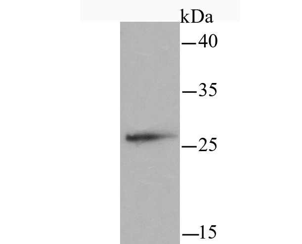
Fig1: Western blot analysis of mtTFA on Hela cell using anti-mtTFA antibody at 1/1,000 dilution.Application
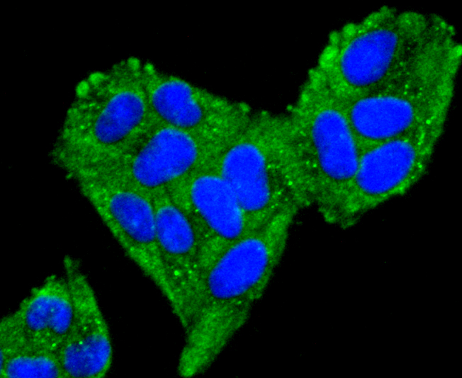
Fig2: ICC staining mtTFA in Hela cells (green). The nuclear counter stain is DAPI (blue). Cells were fixed in paraformaldehyde, permeabilised with 0.25% Triton X100/PBS.Application
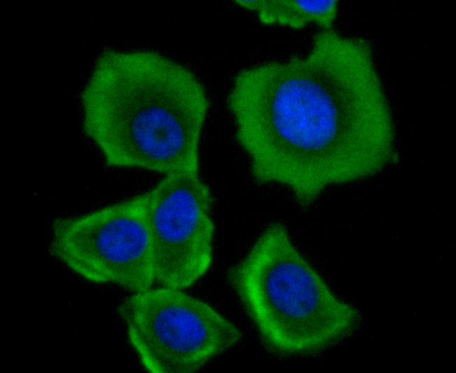
Fig3: ICC staining mtTFA in MCF-7 cells (green). The nuclear counter stain is DAPI (blue). Cells were fixed in paraformaldehyde, permeabilised with 0.25% Triton X100/PBS.Application
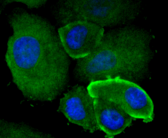
Fig4: ICC staining mtTFA in SK-Br-3 cells (green). The nuclear counter stain is DAPI (blue). Cells were fixed in paraformaldehyde, permeabilised with 0.25% Triton X100/PBS.Application
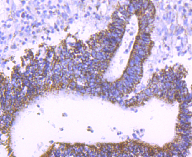
Fig5: Immunohistochemical analysis of paraffin-embedded human colon cancer tissue using anti-mtTFA antibody. Counter stained with hematoxylin.Application
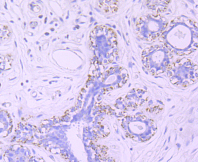
Fig6: Immunohistochemical analysis of paraffin-embedded human breast tissue using anti-mtTFA antibody. Counter stained with hematoxylin.Application
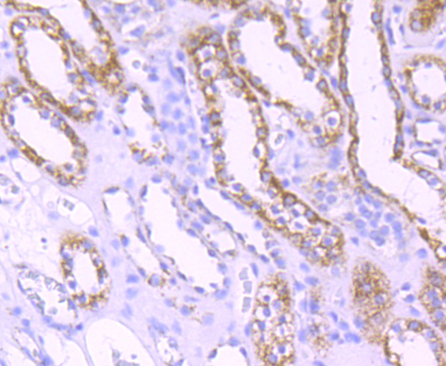
Fig7: Immunohistochemical analysis of paraffin-embedded human kidney tissue using anti-mtTFA antibody. Counter stained with hematoxylin.| Product Name | mtTFA |
|---|---|
| Antibody Type | Primary Antibodies |
| Antigen Alias | anscription factor 6-like 1 antibody Mitochondrial transcription factor 1 antibody mitochondrial transcription factor A antibody MtTF1 antibody mtTFA antibody TCF 6 antibody TCF-6 antibody TCF6 antibody TCF6L1 antibody TCF6L2 antibody TCF6L3 antibody TFAM antibody TFAM_HUMAN antibody Transcription factor 6 antibody Transcription factor 6 like 2 (mitochondrial transcription factor) antibody Transcription factor 6 like 2 antibody Transcription factor 6-like 2 antibody transcription factor 6-like 3 antibody Transcription factor A, mitochondrial antibody Transcription factor A, mitochondrial antibody Transcription factor A, mitochondrial precursor antibody |
| Product description | Binds to the mitochondrial light strand promoter and functions in mitochondrial transcription regulation. Required for accurate and efficient promoter recognition by the mitochondrial RNA polymerase. Promotes transcription initiation from the HSP1 and the light strand promoter by binding immediately upstream of transcriptional start sites. Is able to unwind and bend DNA. Required for maintenance of normal levels of mitochondrial DNA. May play a role in organizing and compacting mitochondrial DNA. target DNA. Interacts with TFB1M and TFB2M. |
| Immunogen | Recombinant protein |
| Clonality | Monoclonal |
|---|---|
| Isotype | IgG |
| Host Species | Recombinant rabbit |
| Tested Applications | WBICC/IFIHCIP |
| WB:1:200-1:500 ICC/IF:1:50-1:200 IHC:1:50-1:200 IP:1:10-1:50 |
|
| Species Reactivity | Human |
| Concentration | 1mg/ml |
| Alternative Names | anscription factor 6-like 1 antibody
Mitochondrial transcription factor 1 antibody
mitochondrial transcription factor A antibody
MtTF1 antibody
mtTFA antibody
TCF 6 antibody
TCF-6 antibody
TCF6 antibody
TCF6L1 antibody
TCF6L2 antibody
TCF6L3 antibody
TFAM antibody
TFAM_HUMAN antibody
Transcription factor 6 antibody
Transcription factor 6 like 2 (mitochondrial transcription factor) antibody
Transcription factor 6 like 2 antibody
Transcription factor 6-like 2 antibody
transcription factor 6-like 3 antibody
Transcription factor A mitochondrial antibody Transcription factor A mitochondrial antibody Transcription factor A mitochondrial precursor antibody |
|---|---|
| Cellular Localization | Mitochondrion. |
| SwissProt ID | Q00059 |
|---|
Application
Fig1: Western blot analysis of mtTFA on Hela cell using anti-mtTFA antibody at 1/1,000 dilution.
Application
Fig2: ICC staining mtTFA in Hela cells (green). The nuclear counter stain is DAPI (blue). Cells were fixed in paraformaldehyde, permeabilised with 0.25% Triton X100/PBS.
Application
Fig3: ICC staining mtTFA in MCF-7 cells (green). The nuclear counter stain is DAPI (blue). Cells were fixed in paraformaldehyde, permeabilised with 0.25% Triton X100/PBS.
Application
Fig4: ICC staining mtTFA in SK-Br-3 cells (green). The nuclear counter stain is DAPI (blue). Cells were fixed in paraformaldehyde, permeabilised with 0.25% Triton X100/PBS.
Application
Fig5: Immunohistochemical analysis of paraffin-embedded human colon cancer tissue using anti-mtTFA antibody. Counter stained with hematoxylin.
Application
Fig6: Immunohistochemical analysis of paraffin-embedded human breast tissue using anti-mtTFA antibody. Counter stained with hematoxylin.
Application
Fig7: Immunohistochemical analysis of paraffin-embedded human kidney tissue using anti-mtTFA antibody. Counter stained with hematoxylin.| Positive Control | Hela, MCF-7, SK-Br-3, human colon cancer tissue, human breast tissue, human kidney tissue. |
|---|---|
| Application Notes | WB:1:200-1:500 ICC/IF:1:50-1:200 IHC:1:50-1:200 IP:1:10-1:50 |
| Form | Liquid |
|---|---|
| Storage Instructions | Store at +4℃ after thawing. Aliquot store at -20℃ or -80℃. Avoid repeated freeze / thaw cycles. |
| Storage Buffer | 1*TBS (pH7.4), 1%BSA, 40%Glycerol. Preservative: 0.05% Sodium Azide. |


