Application
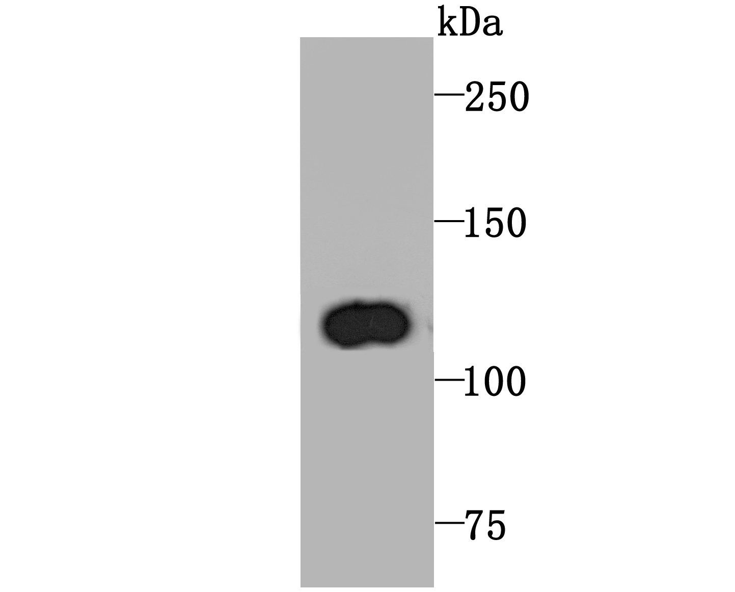
Fig1: Western blot analysis of PGC1 beta on rat spleen tissue lysate using anti-PGC1 beta antibody at 1/1,000 dilution.Application
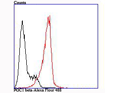
Fig2: Flow cytometric analysis of HL-60 cells with PGC1 beta antibody at 1/100 dilution (red) compared with an unlabelled control (cells without incubation with primary antibody; black).| Product Name | PGC1 beta |
|---|---|
| Antibody Type | Primary Antibodies |
| Antigen Alias | PERC antibody peroxisome proliferative activated receptor, gamma, coactivator 1, antibody peroxisome proliferator-activated receptor gamma coactivator 1 beta antibody Peroxisome proliferator-activated receptor gamma coactivator 1-beta antibody peroxisome proliferator-activated receptor gamma, coactivator 1 beta antibody PGC-1(beta) antibody PGC-1-beta antibody PGC-1-related estrogen receptor alpha coactivator antibody PGC1 antibody PPAR gamma coactivator-1beta antibody PPAR-gamma coactivator 1-beta antibody PPARGC-1-beta antibody PPARGC1 antibody Ppargc1b antibody PRGC2_HUMAN antibody |
| Product description | Plays a role of stimulator of transcription factors and nuclear receptors activities. Activates transcritional activity of estrogen receptor alpha, nuclear respiratory factor 1 (NRF1) and glucocorticoid receptor in the presence of glucocorticoids. May play a role in constitutive non-adrenergic-mediated mitochondrial biogenesis as suggested by increased basal oxygen consumption and mitochondrial number when overexpressed. May be involved in fat oxidation and non-oxidative glucose metabolism and in the regulation of energy expenditure. Induces the expression of PERM1 in the skeletal muscle in an ESRRA-dependent manner. |
| Immunogen | Recombinant protein |
| Clonality | Monoclonal |
|---|---|
| Isotype | IgG |
| Host Species | Recombinant rabbit |
| Tested Applications | WBIPFC |
| WB:1:500-1:2,000 FC:1:50-1:200 IP:1:10-1:50 |
|
| Species Reactivity | HumanRat |
| Concentration | 1mg/ml |
| Alternative Names | PERC antibody
peroxisome proliferative activated receptor gamma coactivator 1 antibody peroxisome proliferator-activated receptor gamma coactivator 1 beta antibody Peroxisome proliferator-activated receptor gamma coactivator 1-beta antibody peroxisome proliferator-activated receptor gamma coactivator 1 beta antibody PGC-1(beta) antibody PGC-1-beta antibody PGC-1-related estrogen receptor alpha coactivator antibody PGC1 antibody PPAR gamma coactivator-1beta antibody PPAR-gamma coactivator 1-beta antibody PPARGC-1-beta antibody PPARGC1 antibody Ppargc1b antibody PRGC2_HUMAN antibody |
|---|---|
| Cellular Localization | Nucleus. |
Application
Fig1: Western blot analysis of PGC1 beta on rat spleen tissue lysate using anti-PGC1 beta antibody at 1/1,000 dilution.
Application
Fig2: Flow cytometric analysis of HL-60 cells with PGC1 beta antibody at 1/100 dilution (red) compared with an unlabelled control (cells without incubation with primary antibody; black).| Positive Control | Rat spleen tissue lysate, HL-60. |
|---|---|
| Application Notes | WB:1:500-1:2,000 FC:1:50-1:200 IP:1:10-1:50 |
| Form | Liquid |
|---|---|
| Storage Instructions | Store at +4℃ after thawing. Aliquot store at -20℃ or -80℃. Avoid repeated freeze / thaw cycles. |
| Storage Buffer | 1*TBS (pH7.4), 1%BSA, 40%Glycerol. Preservative: 0.05% Sodium Azide. |


