Application
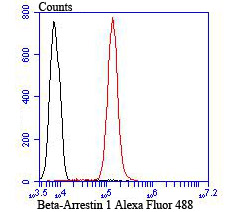
Fig9: Flow cytometric analysis of Jurkat cells with beta Arrestin 1 antibody at 1/100 dilution (red) compared with an unlabelled control (cells without incubation with primary antibody; black). Alexa Fluor 488-conjugated goat anti-rabbit IgG was used as the secondary antibody.Application
Fig9: Flow cytometric analysis of Jurkat cells with beta Arrestin 1 antibody at 1/100 dilution (red) compared with an unlabelled control (cells without incubation with primary antibody; black). Alexa Fluor 488-conjugated goat anti-rabbit IgG was used as the secondary antibody.Application
Fig9: Flow cytometric analysis of Jurkat cells with beta Arrestin 1 antibody at 1/100 dilution (red) compared with an unlabelled control (cells without incubation with primary antibody; black). Alexa Fluor 488-conjugated goat anti-rabbit IgG was used as the secondary antibody.Application
Fig9: Flow cytometric analysis of Jurkat cells with beta Arrestin 1 antibody at 1/100 dilution (red) compared with an unlabelled control (cells without incubation with primary antibody; black). Alexa Fluor 488-conjugated goat anti-rabbit IgG was used as the secondary antibody.Application
Fig9: Flow cytometric analysis of Jurkat cells with beta Arrestin 1 antibody at 1/100 dilution (red) compared with an unlabelled control (cells without incubation with primary antibody; black). Alexa Fluor 488-conjugated goat anti-rabbit IgG was used as the secondary antibody.Application
Fig9: Flow cytometric analysis of Jurkat cells with beta Arrestin 1 antibody at 1/100 dilution (red) compared with an unlabelled control (cells without incubation with primary antibody; black). Alexa Fluor 488-conjugated goat anti-rabbit IgG was used as the secondary antibody.Application
Fig9: Flow cytometric analysis of Jurkat cells with beta Arrestin 1 antibody at 1/100 dilution (red) compared with an unlabelled control (cells without incubation with primary antibody; black). Alexa Fluor 488-conjugated goat anti-rabbit IgG was used as the secondary antibody.Application
Fig9: Flow cytometric analysis of Jurkat cells with beta Arrestin 1 antibody at 1/100 dilution (red) compared with an unlabelled control (cells without incubation with primary antibody; black). Alexa Fluor 488-conjugated goat anti-rabbit IgG was used as the secondary antibody.Application
Fig9: Flow cytometric analysis of Jurkat cells with beta Arrestin 1 antibody at 1/100 dilution (red) compared with an unlabelled control (cells without incubation with primary antibody; black). Alexa Fluor 488-conjugated goat anti-rabbit IgG was used as the secondary antibody.| Product Name | Beta-Arrestin 1 |
|---|---|
| Antibody Type | Primary Antibodies |
| Antigen Alias | ARB1 antibody ARR1 antibody ARRB1 antibody ARRB1_HUMAN antibody Arrestin 2 antibody Arrestin beta 1 antibody Arrestin beta-1 antibody Beta-arrestin-1 antibody |
| Product description | The members of the G protein coupled receptor family are distinguished by their slow transmitting response to ligand binding. These seven transmembrane proteins include the adrenergic, serotonin and dopamine receptors. The effect of the signaling molecule can be excitatory or inhibitory depending on the type of receptor to which it binds. Members of the β-Arrestin family regulate receptor binding to G proteins. β-Arrestins have been found to be located at postsynaptic sites, where they are thought to act in concert with βARK (βARK1, also designated GRK 2, or βARK2, also designated GRK 3) to regulate G protein-coupled neurotransmitter receptors. Expression of β-Arrestin-1 and b-Arrestin-2 is seen predominantly in spleen and neuronal tissues. It has been shown that β-Arrestin-1 expression is modulated by intracellular cAMP, which may be a novel mechanism for the regulation of receptor-mediated responses. |
| Immunogen | Peptide |
| Clonality | Polyclonal |
|---|---|
| Isotype | IgG |
| Host Species | Rabbit |
| Tested Applications | WBICCIHCFC |
| WB:1:500-1:1000 ICC:1:50-1:200 IHC:1:50-1:100 FC:1:50-1:100 |
|
| Species Reactivity | HumanMouseRat |
| Concentration | 1mg/ml |
| Alternative Names | ARB1 antibody ARR1 antibody ARRB1 antibody ARRB1_HUMAN antibody Arrestin 2 antibody Arrestin beta 1 antibody Arrestin beta-1 antibody Beta-arrestin-1 antibody |
|---|---|
| Molecular Weight(MW) | 50 kDa |
| Cellular Localization | Cytoplasm. Membrane. Nucleus. |
Application
Fig9: Flow cytometric analysis of Jurkat cells with beta Arrestin 1 antibody at 1/100 dilution (red) compared with an unlabelled control (cells without incubation with primary antibody; black). Alexa Fluor 488-conjugated goat anti-rabbit IgG was used as the secondary antibody.
Application
Fig9: Flow cytometric analysis of Jurkat cells with beta Arrestin 1 antibody at 1/100 dilution (red) compared with an unlabelled control (cells without incubation with primary antibody; black). Alexa Fluor 488-conjugated goat anti-rabbit IgG was used as the secondary antibody.
Application
Fig9: Flow cytometric analysis of Jurkat cells with beta Arrestin 1 antibody at 1/100 dilution (red) compared with an unlabelled control (cells without incubation with primary antibody; black). Alexa Fluor 488-conjugated goat anti-rabbit IgG was used as the secondary antibody.
Application
Fig9: Flow cytometric analysis of Jurkat cells with beta Arrestin 1 antibody at 1/100 dilution (red) compared with an unlabelled control (cells without incubation with primary antibody; black). Alexa Fluor 488-conjugated goat anti-rabbit IgG was used as the secondary antibody.
Application
Fig9: Flow cytometric analysis of Jurkat cells with beta Arrestin 1 antibody at 1/100 dilution (red) compared with an unlabelled control (cells without incubation with primary antibody; black). Alexa Fluor 488-conjugated goat anti-rabbit IgG was used as the secondary antibody.
Application
Fig9: Flow cytometric analysis of Jurkat cells with beta Arrestin 1 antibody at 1/100 dilution (red) compared with an unlabelled control (cells without incubation with primary antibody; black). Alexa Fluor 488-conjugated goat anti-rabbit IgG was used as the secondary antibody.
Application
Fig9: Flow cytometric analysis of Jurkat cells with beta Arrestin 1 antibody at 1/100 dilution (red) compared with an unlabelled control (cells without incubation with primary antibody; black). Alexa Fluor 488-conjugated goat anti-rabbit IgG was used as the secondary antibody.
Application
Fig9: Flow cytometric analysis of Jurkat cells with beta Arrestin 1 antibody at 1/100 dilution (red) compared with an unlabelled control (cells without incubation with primary antibody; black). Alexa Fluor 488-conjugated goat anti-rabbit IgG was used as the secondary antibody.
Application
Fig9: Flow cytometric analysis of Jurkat cells with beta Arrestin 1 antibody at 1/100 dilution (red) compared with an unlabelled control (cells without incubation with primary antibody; black). Alexa Fluor 488-conjugated goat anti-rabbit IgG was used as the secondary antibody.| Positive Control | Human placenta tissue lysate, A549, HUVEC, LOVO, rat brain tissue, human colon cancer tissue, human breast tissue, mouse colon tissue, Jurkat. |
|---|---|
| Application Notes | WB:1:500-1:1000 ICC:1:50-1:200 IHC:1:50-1:100 FC:1:50-1:100 |
| Form | Liquid |
|---|---|
| Storage Instructions | Store at +4℃ after thawing. Aliquot store at -20℃ or -80℃. Avoid repeated freeze / thaw cycles. |
| Storage Buffer | 1*TBS (pH7.4), 0.5%BSA, 50%Glycerol. Preservative: 0.05% Sodium Azide. |


