Application
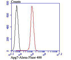
Fig7: Flow cytometric analysis of Jurkat cells with Apg7 antibody at 1/100 dilution (red) compared with an unlabelled control (cells without incubation with primary antibody; black).Application
Fig7: Flow cytometric analysis of Jurkat cells with Apg7 antibody at 1/100 dilution (red) compared with an unlabelled control (cells without incubation with primary antibody; black).Application
Fig7: Flow cytometric analysis of Jurkat cells with Apg7 antibody at 1/100 dilution (red) compared with an unlabelled control (cells without incubation with primary antibody; black).Application
Fig7: Flow cytometric analysis of Jurkat cells with Apg7 antibody at 1/100 dilution (red) compared with an unlabelled control (cells without incubation with primary antibody; black).Application
Fig7: Flow cytometric analysis of Jurkat cells with Apg7 antibody at 1/100 dilution (red) compared with an unlabelled control (cells without incubation with primary antibody; black).Application
Fig7: Flow cytometric analysis of Jurkat cells with Apg7 antibody at 1/100 dilution (red) compared with an unlabelled control (cells without incubation with primary antibody; black).Application
Fig7: Flow cytometric analysis of Jurkat cells with Apg7 antibody at 1/100 dilution (red) compared with an unlabelled control (cells without incubation with primary antibody; black).| Product Name | Apg7 |
|---|---|
| Antibody Type | Primary Antibodies |
| Antigen Alias | 1810013K23Rik antibody Apg 7 antibody APG7 autophagy 7 like antibody APG7 autophagy 7-like (S. cerevisiae) antibody APG7 like antibody APG7, S. cerevisiae, homolog of antibody APG7-like antibody APG7L antibody ATG 7 antibody ATG12-activating enzyme E1 ATG7 antibody ATG7 antibody ATG7 autophagy related 7 homolog (S. cerevisiae) antibody ATG7 autophagy related 7 homolog antibody ATG7_HUMAN antibody Atg7l antibody Autophagy 7, S. cerevisiae, homolog of antibody Autophagy related protein 7 antibody Autophagy-related 7 (yeast) antibody Autophagy-related protein 7 antibody DKFZp434N0735 antibody GSA 7 antibody GSA7 antibody hAGP7 antibody Ubiquitin activating enzyme E1 like protein antibody Ubiquitin-activating enzyme E1-like protein antibody Ubiquitin-like modifier-activating enzyme ATG7 antibody |
| Product description | In yeast, autophagy is an essential process for survival during nutrient starvation and cell differentiation. The process of autophagy is characterized as a non-selective degradation of cytoplasmic proteins into membrane stuctures called autophagosomes, and it is dependent on several proteins, including the autophagy proteins APG5 and APG7. Yeast Apg7 and the human homolog, APG7, share similarities with the ubiquitin-activating enzyme E1 in Saccharomyces cerevisiae, and are likewise responsible for enzymatically activating the autophagy conjugation system. Apg5 and the human homolog, APG5 (also designated apoptosis specific protein or APS), function as substrates for the autophagy protein APG12. These proteins are covalently bonded together to form APG12/APG5 conjugates, which are required for the progression of autophagy. |
| Immunogen | Peptide |
| Clonality | Polyclonal |
|---|---|
| Isotype | IgG |
| Host Species | Rabbit |
| Tested Applications | ICCIHCFC |
| ICC:1:500-1:2,000 IHC:1:200-1:1,000 FC:1:50-1:100 |
|
| Species Reactivity | HumanMouse |
| Concentration | 1mg/ml |
| Alternative Names | 1810013K23Rik antibody
Apg 7 antibody
APG7 autophagy 7 like antibody
APG7 autophagy 7-like (S. cerevisiae) antibody
APG7 like antibody
APG7 S. cerevisiae homolog of antibody APG7-like antibody APG7L antibody ATG 7 antibody ATG12-activating enzyme E1 ATG7 antibody ATG7 antibody ATG7 autophagy related 7 homolog (S. cerevisiae) antibody ATG7 autophagy related 7 homolog antibody ATG7_HUMAN antibody Atg7l antibody Autophagy 7 S. cerevisiae homolog of antibody Autophagy related protein 7 antibody Autophagy-related 7 (yeast) antibody Autophagy-related protein 7 antibody DKFZp434N0735 antibody GSA 7 antibody GSA7 antibody hAGP7 antibody Ubiquitin activating enzyme E1 like protein antibody Ubiquitin-activating enzyme E1-like protein antibody Ubiquitin-like modifier-activating enzyme ATG7 antibody |
|---|---|
| Molecular Weight(MW) | 78 kDa |
| Cellular Localization | Cytoplasm. |
Application
Fig7: Flow cytometric analysis of Jurkat cells with Apg7 antibody at 1/100 dilution (red) compared with an unlabelled control (cells without incubation with primary antibody; black).
Application
Fig7: Flow cytometric analysis of Jurkat cells with Apg7 antibody at 1/100 dilution (red) compared with an unlabelled control (cells without incubation with primary antibody; black).
Application
Fig7: Flow cytometric analysis of Jurkat cells with Apg7 antibody at 1/100 dilution (red) compared with an unlabelled control (cells without incubation with primary antibody; black).
Application
Fig7: Flow cytometric analysis of Jurkat cells with Apg7 antibody at 1/100 dilution (red) compared with an unlabelled control (cells without incubation with primary antibody; black).
Application
Fig7: Flow cytometric analysis of Jurkat cells with Apg7 antibody at 1/100 dilution (red) compared with an unlabelled control (cells without incubation with primary antibody; black).
Application
Fig7: Flow cytometric analysis of Jurkat cells with Apg7 antibody at 1/100 dilution (red) compared with an unlabelled control (cells without incubation with primary antibody; black).
Application
Fig7: Flow cytometric analysis of Jurkat cells with Apg7 antibody at 1/100 dilution (red) compared with an unlabelled control (cells without incubation with primary antibody; black).| Positive Control | A431, A549, HUVEC, human breast tissue, human placenta tissue, mouse kidney tissue, Jurkat. |
|---|---|
| Application Notes | ICC:1:500-1:2,000 IHC:1:200-1:1,000 FC:1:50-1:100 |
| Form | Liquid |
|---|---|
| Storage Instructions | Store at +4℃ after thawing. Aliquot store at -20℃ or -80℃. Avoid repeated freeze / thaw cycles. |
| Storage Buffer | 1*TBS (pH7.4), 0.5%BSA, 50%Glycerol. Preservative: 0.05% Sodium Azide. |


