Application
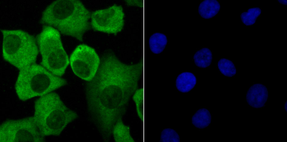
Fig1: ICC staining Progesterone Receptor in A549 cells (green). The nuclear counter stain is DAPI (blue). Cells were fixed in paraformaldehyde, permeabilised with 0.25% Triton X100/PBS.Application
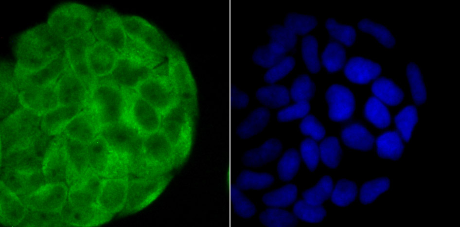
Fig2: ICC staining Progesterone Receptor in Hela cells (green). The nuclear counter stain is DAPI (blue). Cells were fixed in paraformaldehyde, permeabilised with 0.25% Triton X100/PBS.Application
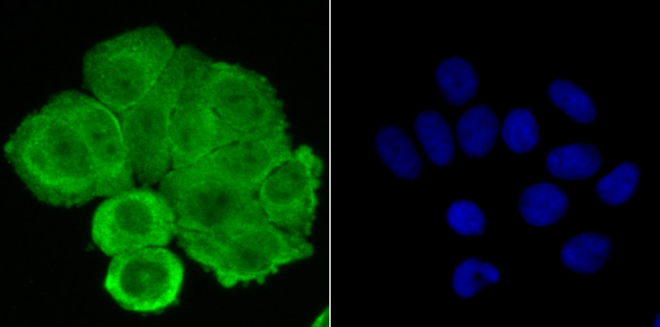
Fig3: ICC staining Progesterone Receptor in MCF-7 cells (green). The nuclear counter stain is DAPI (blue). Cells were fixed in paraformaldehyde, permeabilised with 0.25% Triton X100/PBS.Application
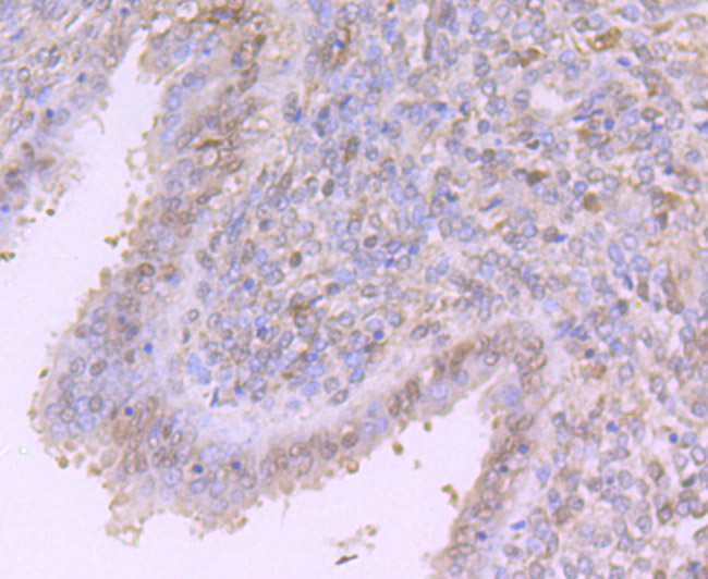
Fig4: Immunohistochemical analysis of paraffin-embedded human uterus tissue using anti-Progesterone Receptor antibody. Counter stained with hematoxylin.Application
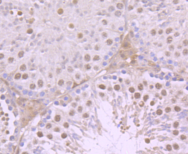
Fig5: Immunohistochemical analysis of paraffin-embedded mouse testis tissue using anti-Progesterone Receptor antibody. Counter stained with hematoxylin.| Product Name | Progesterone Receptor |
|---|---|
| Antibody Type | Primary Antibodies |
| Antigen Alias | NR3C3 antibody Nuclear receptor subfamily 3 group C member 3 antibody PGR antibody PR antibody PRA antibody PRB antibody PRGR_HUMAN antibody Progesterone receptor antibody Progestin receptor form A antibody Progestin receptor form B antibody |
| Product description | The effects of progesterone are mediated by two functionally different isoforms of the progesterone receptor, PR-A and PR-B, which are transcribed from distinct, estrogen-inducible promoters within a single copy of the PR gene. The first 164 amino acids of PR-B are absent in PR-A. Progesterone-bound PR-A and PR-B have different transcription activation properties. Specifically, PR-B functions as a transcriptional activator in most cell and promoter contexts, while PR-A is transcriptionally inactive and functions as a strong ligand-dependent transdominant repressor of steroid hormone receptor transcriptional activity. An inhibitory domain (ID), which maps to the amino terminus of the receptor, exists within both PR isoforms. Interestingly, the ID is functionally active only in PR-A and is necessary for steroid hormone transrepression by PR-A, suggesting that PR-A and PR-B may have different conformations in the cell. |
| Immunogen | Peptide |
| Clonality | Polyclonal |
|---|---|
| Isotype | IgG |
| Host Species | Rabbit |
| Tested Applications | ICCIHC |
| ICC:1:500-1:1,000 IHC:1:100-1:500 |
|
| Species Reactivity | HumanMouse |
| Concentration | 1mg/ml |
| Alternative Names | NR3C3 antibody Nuclear receptor subfamily 3 group C member 3 antibody PGR antibody PR antibody PRA antibody PRB antibody PRGR_HUMAN antibody Progesterone receptor antibody Progestin receptor form A antibody Progestin receptor form B antibody |
|---|---|
| Molecular Weight(MW) | 99 kDa |
| Cellular Localization | Cytoplasm. Nucleus. |
Application
Fig1: ICC staining Progesterone Receptor in A549 cells (green). The nuclear counter stain is DAPI (blue). Cells were fixed in paraformaldehyde, permeabilised with 0.25% Triton X100/PBS.
Application
Fig2: ICC staining Progesterone Receptor in Hela cells (green). The nuclear counter stain is DAPI (blue). Cells were fixed in paraformaldehyde, permeabilised with 0.25% Triton X100/PBS.
Application
Fig3: ICC staining Progesterone Receptor in MCF-7 cells (green). The nuclear counter stain is DAPI (blue). Cells were fixed in paraformaldehyde, permeabilised with 0.25% Triton X100/PBS.
Application
Fig4: Immunohistochemical analysis of paraffin-embedded human uterus tissue using anti-Progesterone Receptor antibody. Counter stained with hematoxylin.
Application
Fig5: Immunohistochemical analysis of paraffin-embedded mouse testis tissue using anti-Progesterone Receptor antibody. Counter stained with hematoxylin.| Positive Control | A549, Hela, MCF-7, human uterus tissue, mouse testis tissue. |
|---|---|
| Application Notes | ICC:1:500-1:1,000 IHC:1:100-1:500 |
| Form | Liquid |
|---|---|
| Storage Instructions | Store at +4℃ after thawing. Aliquot store at -20℃ or -80℃. Avoid repeated freeze / thaw cycles. |
| Storage Buffer | 1*TBS (pH7.4), 0.5%BSA, 50%Glycerol. Preservative: 0.05% Sodium Azide. |


