Application
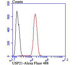
Fig7: Flow cytometric analysis of SH-SY-5Y cells with USP21 antibody at 1/100 dilution (red) compared with an unlabelled control (cells without incubation with primary antibody; black). Alexa Fluor 488-conjugated goat anti-rabbit IgG was used as the secondary antibody.Application
Fig7: Flow cytometric analysis of SH-SY-5Y cells with USP21 antibody at 1/100 dilution (red) compared with an unlabelled control (cells without incubation with primary antibody; black). Alexa Fluor 488-conjugated goat anti-rabbit IgG was used as the secondary antibody.Application
Fig7: Flow cytometric analysis of SH-SY-5Y cells with USP21 antibody at 1/100 dilution (red) compared with an unlabelled control (cells without incubation with primary antibody; black). Alexa Fluor 488-conjugated goat anti-rabbit IgG was used as the secondary antibody.Application
Fig7: Flow cytometric analysis of SH-SY-5Y cells with USP21 antibody at 1/100 dilution (red) compared with an unlabelled control (cells without incubation with primary antibody; black). Alexa Fluor 488-conjugated goat anti-rabbit IgG was used as the secondary antibody.Application
Fig7: Flow cytometric analysis of SH-SY-5Y cells with USP21 antibody at 1/100 dilution (red) compared with an unlabelled control (cells without incubation with primary antibody; black). Alexa Fluor 488-conjugated goat anti-rabbit IgG was used as the secondary antibody.Application
Fig7: Flow cytometric analysis of SH-SY-5Y cells with USP21 antibody at 1/100 dilution (red) compared with an unlabelled control (cells without incubation with primary antibody; black). Alexa Fluor 488-conjugated goat anti-rabbit IgG was used as the secondary antibody.Application
Fig7: Flow cytometric analysis of SH-SY-5Y cells with USP21 antibody at 1/100 dilution (red) compared with an unlabelled control (cells without incubation with primary antibody; black). Alexa Fluor 488-conjugated goat anti-rabbit IgG was used as the secondary antibody.| Product Name | USP21 |
|---|---|
| Antibody Type | Primary Antibodies |
| Antigen Alias | Deubiquitinating enzyme 21 antibody deubiquitinating enzyme 23 antibody EC 3.1.2.15 antibody ESTM28 antibody MGC3394 antibody NEDD8-specific protease antibody Ubiquitin carboxyl terminal hydrolase 21 antibody ubiquitin carboxyl terminal hydrolase 23 antibody Ubiquitin carboxyl-terminal hydrolase 21 antibody ubiquitin specific peptidase 21 antibody Ubiquitin specific processing protease 21 antibody ubiquitin specific protease 16 antibody ubiquitin specific protease 21 antibody ubiquitin specific protease 23 antibody Ubiquitin thioesterase 21 antibody Ubiquitin thiolesterase 21 antibody ubiquitin thiolesterase 23 antibody ubiquitin-specific protease 23, formerly antibody Ubiquitin-specific-processing protease 21 antibody UBP21_HUMAN antibody USP16 antibodyv Usp21 antibody USP23 antibody USP23, formerly antibody W53272 antibody |
| Product description | The ubiquitin (Ub) pathway involves three sequential enzymatic steps that facilitate the conjugation of Ub and Ub-like molecules to specific protein substrates. Through the use of a wide range of enzymes that can add or remove ubiquitin, the Ub pathway controls many intracellular processes such as signal transduction, transcriptional activation and cell cycle progression. USP21 (ubiquitin specific peptidase 21), also known as USP16 or USP23, is a 565 amino acid protein that belongs to the C19 peptidase family of ubiquitin carboxy-terminal hydrolases. Capable of removing ubiquitin from ubiquitinated proteins, USP21 plays a role in signal transduction and can also remove NEDD8 from NEDD8-conjugated proteins, possibly functioning to influence NEDD8-mediated protein proteolysis. Multiple isoforms of USP21 exist due to alternative splicing events. |
| Immunogen | Peptide |
| Clonality | Polyclonal |
|---|---|
| Isotype | IgG |
| Host Species | Rabbit |
| Tested Applications | WBICCIHCFC |
| WB:1:500 ICC:1:500-1:2,000 IHC:1:50-1:200 FC:1:50-1:100 |
|
| Species Reactivity | HumanRatMouse |
| Concentration | 1mg/ml |
| Alternative Names | Deubiquitinating enzyme 21 antibody
deubiquitinating enzyme 23 antibody
EC 3.1.2.15 antibody
ESTM28 antibody
MGC3394 antibody
NEDD8-specific protease antibody
Ubiquitin carboxyl terminal hydrolase 21 antibody
ubiquitin carboxyl terminal hydrolase 23 antibody
Ubiquitin carboxyl-terminal hydrolase 21 antibody
ubiquitin specific peptidase 21 antibody
Ubiquitin specific processing protease 21 antibody
ubiquitin specific protease 16 antibody
ubiquitin specific protease 21 antibody
ubiquitin specific protease 23 antibody
Ubiquitin thioesterase 21 antibody
Ubiquitin thiolesterase 21 antibody
ubiquitin thiolesterase 23 antibody
ubiquitin-specific protease 23 formerly antibody Ubiquitin-specific-processing protease 21 antibody UBP21_HUMAN antibody USP16 antibodyv Usp21 antibody USP23 antibody USP23 formerly antibody W53272 antibody |
|---|---|
| Molecular Weight(MW) | 63 kDa |
| Cellular Localization | Cytoplasm. Nucleus. |
Application
Fig7: Flow cytometric analysis of SH-SY-5Y cells with USP21 antibody at 1/100 dilution (red) compared with an unlabelled control (cells without incubation with primary antibody; black). Alexa Fluor 488-conjugated goat anti-rabbit IgG was used as the secondary antibody.
Application
Fig7: Flow cytometric analysis of SH-SY-5Y cells with USP21 antibody at 1/100 dilution (red) compared with an unlabelled control (cells without incubation with primary antibody; black). Alexa Fluor 488-conjugated goat anti-rabbit IgG was used as the secondary antibody.
Application
Fig7: Flow cytometric analysis of SH-SY-5Y cells with USP21 antibody at 1/100 dilution (red) compared with an unlabelled control (cells without incubation with primary antibody; black). Alexa Fluor 488-conjugated goat anti-rabbit IgG was used as the secondary antibody.
Application
Fig7: Flow cytometric analysis of SH-SY-5Y cells with USP21 antibody at 1/100 dilution (red) compared with an unlabelled control (cells without incubation with primary antibody; black). Alexa Fluor 488-conjugated goat anti-rabbit IgG was used as the secondary antibody.
Application
Fig7: Flow cytometric analysis of SH-SY-5Y cells with USP21 antibody at 1/100 dilution (red) compared with an unlabelled control (cells without incubation with primary antibody; black). Alexa Fluor 488-conjugated goat anti-rabbit IgG was used as the secondary antibody.
Application
Fig7: Flow cytometric analysis of SH-SY-5Y cells with USP21 antibody at 1/100 dilution (red) compared with an unlabelled control (cells without incubation with primary antibody; black). Alexa Fluor 488-conjugated goat anti-rabbit IgG was used as the secondary antibody.
Application
Fig7: Flow cytometric analysis of SH-SY-5Y cells with USP21 antibody at 1/100 dilution (red) compared with an unlabelled control (cells without incubation with primary antibody; black). Alexa Fluor 488-conjugated goat anti-rabbit IgG was used as the secondary antibody.| Positive Control | Mouse thymus tissue lysate, PC-3M, SH-SY-5Y, human liver cancer tissue, human kidney tissue, human fetal skeletal muscle tissue. |
|---|---|
| Application Notes | WB:1:500 ICC:1:500-1:2,000 IHC:1:50-1:200 FC:1:50-1:100 |
| Form | Liquid |
|---|---|
| Storage Instructions | Store at +4℃ after thawing. Aliquot store at -20℃ or -80℃. Avoid repeated freeze / thaw cycles. |
| Storage Buffer | 1*TBS (pH7.4), 0.5%BSA, 50%Glycerol. Preservative: 0.05% Sodium Azide. |


