Application
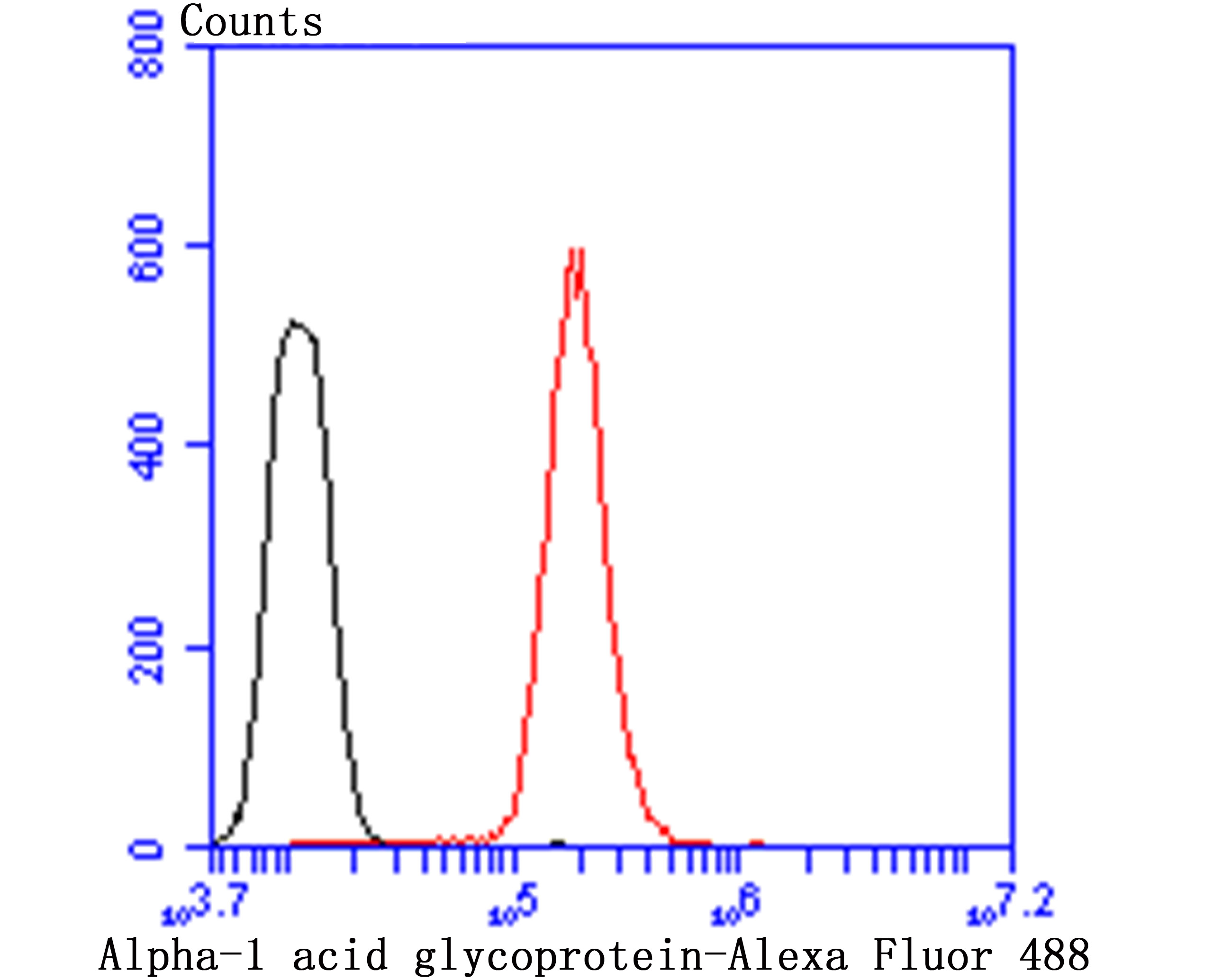
Fig5: Flow cytometric analysis of HepG2 cells with Alpha-1 Acid Glycoprotein antibody at 1/50 dilution (red) compared with an unlabelled control (cells without incubation with primary antibody; black). Alexa Fluor 488-conjugated goat anti-rabbit IgG was used as the secondary antibody.Application
Fig5: Flow cytometric analysis of HepG2 cells with Alpha-1 Acid Glycoprotein antibody at 1/50 dilution (red) compared with an unlabelled control (cells without incubation with primary antibody; black). Alexa Fluor 488-conjugated goat anti-rabbit IgG was used as the secondary antibody.Application
Fig5: Flow cytometric analysis of HepG2 cells with Alpha-1 Acid Glycoprotein antibody at 1/50 dilution (red) compared with an unlabelled control (cells without incubation with primary antibody; black). Alexa Fluor 488-conjugated goat anti-rabbit IgG was used as the secondary antibody.Application
Fig5: Flow cytometric analysis of HepG2 cells with Alpha-1 Acid Glycoprotein antibody at 1/50 dilution (red) compared with an unlabelled control (cells without incubation with primary antibody; black). Alexa Fluor 488-conjugated goat anti-rabbit IgG was used as the secondary antibody.Application
Fig5: Flow cytometric analysis of HepG2 cells with Alpha-1 Acid Glycoprotein antibody at 1/50 dilution (red) compared with an unlabelled control (cells without incubation with primary antibody; black). Alexa Fluor 488-conjugated goat anti-rabbit IgG was used as the secondary antibody.| Product Name | Alpha-1 acid glycoprotein |
|---|---|
| Antibody Type | Primary Antibodies |
| Antigen Alias | A1AG1_HUMAN antibody AGP 1 antibody AGP A antibody AGP antibody AGP1 antibody Alpha 1 acid glycoprotein antibody Alpha-1-acid glycoprotein 1 antibody alpha-1-AGP antibody Epididymis secretory sperm binding protein Li 153w antibody glycoprotein, alpha-1-acid, of serum antibody HEL S 153w antibody OMD 1 antibody ORM antibody ORM1 antibody Orosomucoid 1 antibody Orosomucoid-1 antibody |
| Product description | AGP (α1-acid glycoprotein) is an acute phase plasma protein synthesized by the liver. It functions to regulate the interaction between blood cells and endothelial cells, and together with haptoglobin and C reactive protein, it also mediates the extravasation of cells during infection and inflammation. Expression of AGP is induced by acute-phase stimulatory agents such as bacterial lipopolysaccharides. AGP has a high affinity, low capacity binding for basic drugs at physiological pH. In human plasma, AGP is found at levels of 0.5-1.4 mg/ml, though this is elevated during acute inflammation, and, as a result, levels of this protein can be used to diagnose inflammatory conditions. AGP-1 and AGP-2 contain five and six potential N-glycosylation sites, respectively. Abnormal expression of the APG-1 gene is linked to sarcoidosis and other immunogenetic diseases, while mutations in the APG-2 gene are associated with different types of carcinomas. |
| Immunogen | Recombinant protein |
| Clonality | Polyclonal |
|---|---|
| Isotype | IgG |
| Host Species | Rabbit |
| Tested Applications | WBIHCFC |
| WB:1:500-1:2000 IHC:1:50-1:200 FC:1:50-1:100 |
|
| Species Reactivity | HumanMouseRat |
| Concentration | 1mg/ml |
| Alternative Names | A1AG1_HUMAN antibody
AGP 1 antibody
AGP A antibody
AGP antibody
AGP1 antibody
Alpha 1 acid glycoprotein antibody
Alpha-1-acid glycoprotein 1 antibody
alpha-1-AGP antibody
Epididymis secretory sperm binding protein Li 153w antibody
glycoprotein alpha-1-acid of serum antibody HEL S 153w antibody OMD 1 antibody ORM antibody ORM1 antibody Orosomucoid 1 antibody Orosomucoid-1 antibody |
|---|---|
| Molecular Weight(MW) | 35 kDa |
| Cellular Localization | Secreted. |
Application
Fig5: Flow cytometric analysis of HepG2 cells with Alpha-1 Acid Glycoprotein antibody at 1/50 dilution (red) compared with an unlabelled control (cells without incubation with primary antibody; black). Alexa Fluor 488-conjugated goat anti-rabbit IgG was used as the secondary antibody.
Application
Fig5: Flow cytometric analysis of HepG2 cells with Alpha-1 Acid Glycoprotein antibody at 1/50 dilution (red) compared with an unlabelled control (cells without incubation with primary antibody; black). Alexa Fluor 488-conjugated goat anti-rabbit IgG was used as the secondary antibody.
Application
Fig5: Flow cytometric analysis of HepG2 cells with Alpha-1 Acid Glycoprotein antibody at 1/50 dilution (red) compared with an unlabelled control (cells without incubation with primary antibody; black). Alexa Fluor 488-conjugated goat anti-rabbit IgG was used as the secondary antibody.
Application
Fig5: Flow cytometric analysis of HepG2 cells with Alpha-1 Acid Glycoprotein antibody at 1/50 dilution (red) compared with an unlabelled control (cells without incubation with primary antibody; black). Alexa Fluor 488-conjugated goat anti-rabbit IgG was used as the secondary antibody.
Application
Fig5: Flow cytometric analysis of HepG2 cells with Alpha-1 Acid Glycoprotein antibody at 1/50 dilution (red) compared with an unlabelled control (cells without incubation with primary antibody; black). Alexa Fluor 488-conjugated goat anti-rabbit IgG was used as the secondary antibody.| Positive Control | Human liver tissue lysates, rat liver tissue, human liver tissue, mouse liver tissue, HepG2. |
|---|---|
| Application Notes | WB:1:500-1:2000 IHC:1:50-1:200 FC:1:50-1:100 |
| Form | Liquid |
|---|---|
| Storage Instructions | Store at +4℃ after thawing. Aliquot store at -20℃ or -80℃. Avoid repeated freeze / thaw cycles. |
| Storage Buffer | 1*TBS (pH7.4), 0.5%BSA, 50%Glycerol. Preservative: 0.05% Sodium Azide. |


