Application
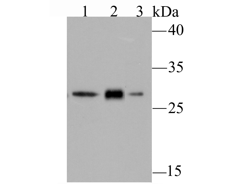
Fig1: Western blot analysis of GCLM on different cell lysates using anti-GCLM antibody at 1/500 dilution. Positive control: Lane 1: A431 Lane 2: PC-12 Lane 3: NIH-3T3Application
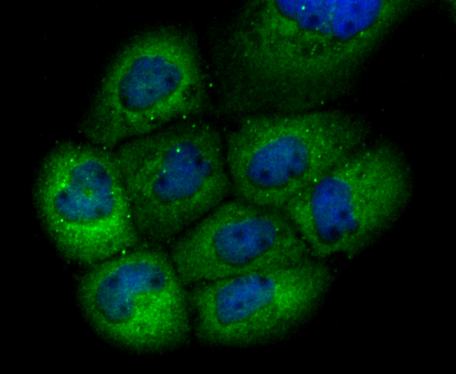
Fig2: ICC staining GCLM in A431 cells (green). The nuclear counter stain is DAPI (blue). Cells were fixed in paraformaldehyde, permeabilised with 0.25% Triton X100/PBS.Application
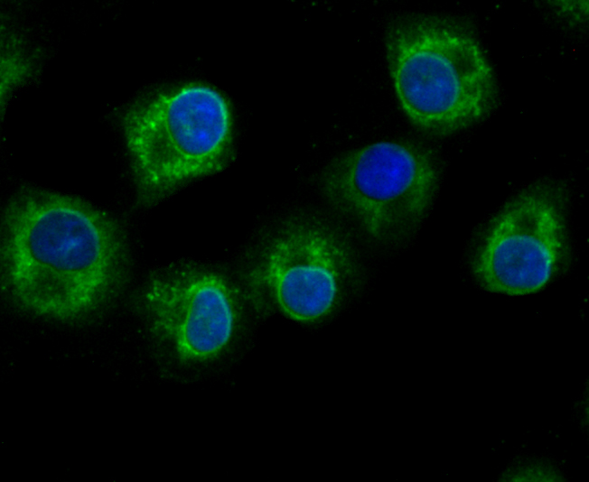
Fig3: ICC staining GCLM in A549 cells (green). The nuclear counter stain is DAPI (blue). Cells were fixed in paraformaldehyde, permeabilised with 0.25% Triton X100/PBS.Application
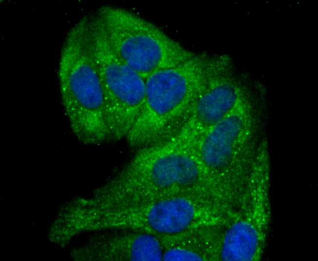
Fig4: ICC staining GCLM in Hela cells (green). The nuclear counter stain is DAPI (blue). Cells were fixed in paraformaldehyde, permeabilised with 0.25% Triton X100/PBS.Application
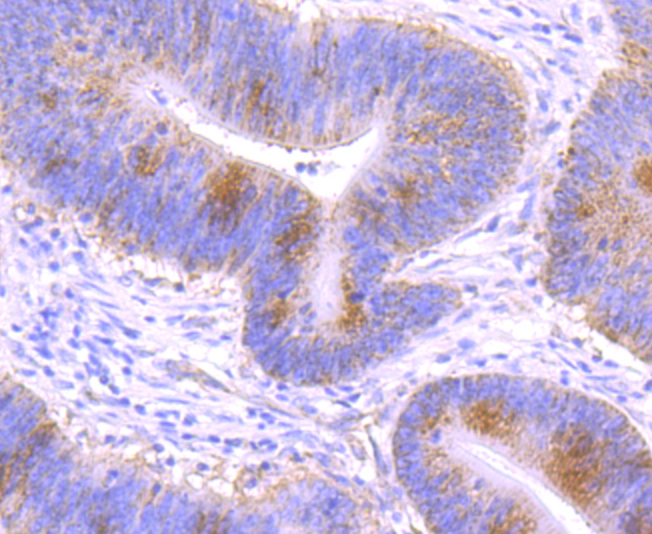
Fig5: Immunohistochemical analysis of paraffin-embedded human colon cancer tissue using anti-GCLM antibody. Counter stained with hematoxylin.Application
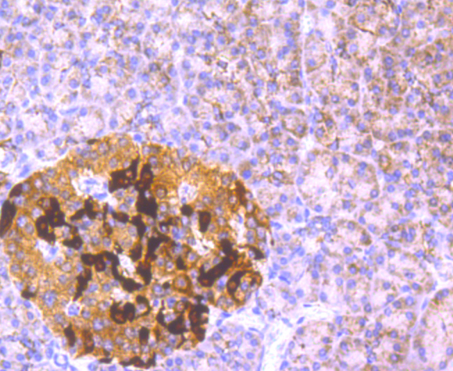
Fig6: Immunohistochemical analysis of paraffin-embedded human pancreas tissue using anti-GCLM antibody. Counter stained with hematoxylin.Application
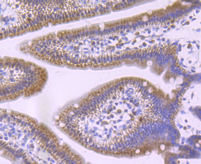
Fig7: Immunohistochemical analysis of paraffin-embedded mouse small intestine tissue using anti-GCLM antibody. Counter stained with hematoxylin.Application
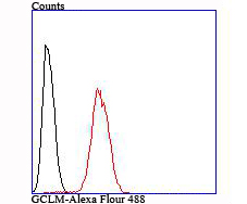
Fig8: Flow cytometric analysis of Hela cells with GCLM antibody at 1/100 dilution (red) compared with an unlabelled control (cells without incubation with primary antibody; black).| Product Name | GCLM |
|---|---|
| Antibody Type | Primary Antibodies |
| Antigen Alias | Gamma ECS regulatory subunit antibody Gamma-ECS regulatory subunit antibody Gamma-glutamylcysteine synthetase regulatory subunit antibody GCLM antibody GCS light chain antibody GLCLR antibody Glutamate cysteine ligase regulatory subunit antibody Glutamatecysteine ligase modifier subunit antibody Glutamatecysteine ligase regulatory subunit antibody GSC light chain antibody GSH0_HUMAN antibody |
| Product description | Gamma-glutamylcysteine synthetase (γ-GCS) is the rate limiting enzyme for glutathione (L-gamma-glutamyl-L-cysteinylglycine, GSH) synthesis. GSH is ubiquitous in mammalian cells as a vital intra- and extracellular protective antioxidant. γ-GCS is a heterodimer of a heavy catalytic subunit and a light regulatory subunit that is responsive to inflammation, phenolic antioxidants, heat shock, oxidants and cytokines. The human gamma-GCS gene encoding the 367 amino acid catalytic subunit maps to chromosome 6p12. The human γ-GCS gene encoding the regulatory subunit maps to chromosome 1p22-p21. The two subunits of γ-GCS form a heterodimeric zinc metalloprotein that gains activity through formation of a reversible disulfide bond. |
| Immunogen | Recombinant protein |
| Clonality | Monoclonal |
|---|---|
| Isotype | IgG |
| Host Species | Recombinant rabbit |
| Tested Applications | WBICC/IFIHCFCIP |
| WB:1:500-1:1,000 ICC:1:50-1:200 IHC:1:50-1:100 FC:1:50-1:100 |
|
| Species Reactivity | HumanMouseRat |
| Concentration | 1mg/ml |
| Alternative Names | Gamma ECS regulatory subunit antibody Gamma-ECS regulatory subunit antibody Gamma-glutamylcysteine synthetase regulatory subunit antibody GCLM antibody GCS light chain antibody GLCLR antibody Glutamate cysteine ligase regulatory subunit antibody Glutamatecysteine ligase modifier subunit antibody Glutamatecysteine ligase regulatory subunit antibody GSC light chain antibody GSH0_HUMAN antibody |
|---|---|
| Cellular Localization | Cytosol. |
Application
Fig1: Western blot analysis of GCLM on different cell lysates using anti-GCLM antibody at 1/500 dilution. Positive control: Lane 1: A431 Lane 2: PC-12 Lane 3: NIH-3T3
Application
Fig2: ICC staining GCLM in A431 cells (green). The nuclear counter stain is DAPI (blue). Cells were fixed in paraformaldehyde, permeabilised with 0.25% Triton X100/PBS.
Application
Fig3: ICC staining GCLM in A549 cells (green). The nuclear counter stain is DAPI (blue). Cells were fixed in paraformaldehyde, permeabilised with 0.25% Triton X100/PBS.
Application
Fig4: ICC staining GCLM in Hela cells (green). The nuclear counter stain is DAPI (blue). Cells were fixed in paraformaldehyde, permeabilised with 0.25% Triton X100/PBS.
Application
Fig5: Immunohistochemical analysis of paraffin-embedded human colon cancer tissue using anti-GCLM antibody. Counter stained with hematoxylin.
Application
Fig6: Immunohistochemical analysis of paraffin-embedded human pancreas tissue using anti-GCLM antibody. Counter stained with hematoxylin.
Application
Fig7: Immunohistochemical analysis of paraffin-embedded mouse small intestine tissue using anti-GCLM antibody. Counter stained with hematoxylin.
Application
Fig8: Flow cytometric analysis of Hela cells with GCLM antibody at 1/100 dilution (red) compared with an unlabelled control (cells without incubation with primary antibody; black).| Positive Control | A431, PC-12, NIH-3T3, Hela, A549,human colon cancer tissue, human pancreas tissue, mouse small intestine tissue. |
|---|---|
| Application Notes | WB:1:500-1:1,000 ICC:1:50-1:200 IHC:1:50-1:100 FC:1:50-1:100 |
| Form | Liquid |
|---|---|
| Storage Instructions | Store at +4℃ after thawing. Aliquot store at -20℃ or -80℃. Avoid repeated freeze / thaw cycles. |
| Storage Buffer | 1*TBS (pH7.4), 1%BSA, 40%Glycerol. Preservative: 0.05% Sodium Azide. |


