Application
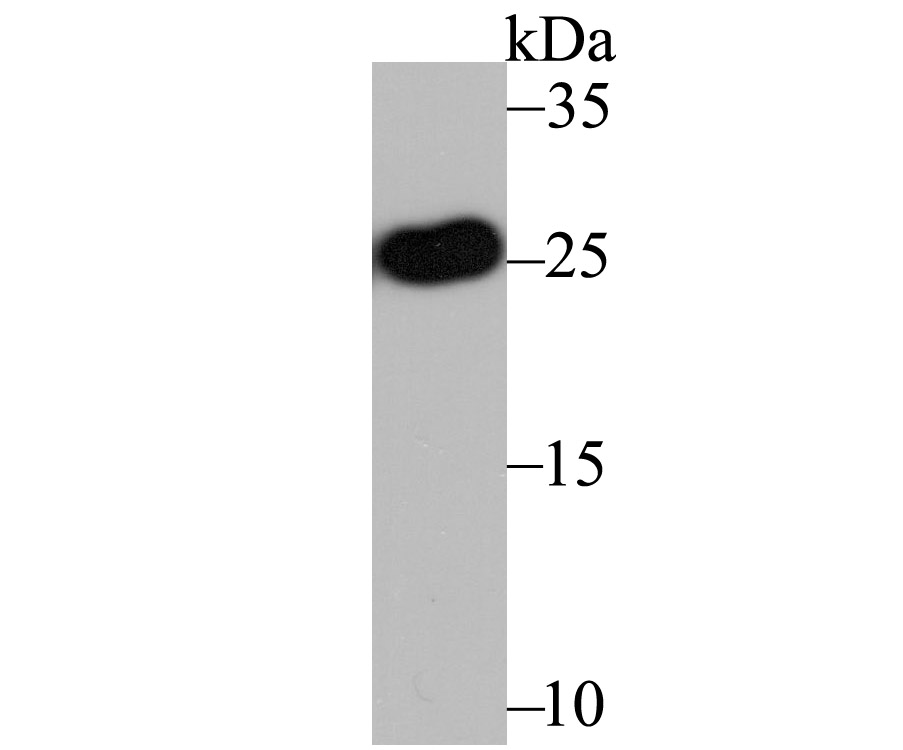
Fig1: Western blot analysis of Apolipoprotein A1 on human liver tissue lysate using anti-Apolipoprotein A1 antibody at 1/1,000 dilution.Application
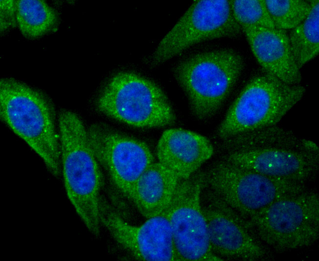
Fig2: ICC staining Apolipoprotein A1 in HepG2 cells (green). The nuclear counter stain is DAPI (blue). Cells were fixed in paraformaldehyde, permeabilised with 0.25% Triton X100/PBS.Application
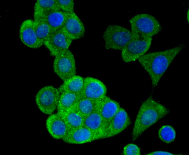
Fig3: ICC staining Apolipoprotein A1 in LOVO cells (green). The nuclear counter stain is DAPI (blue). Cells were fixed in paraformaldehyde, permeabilised with 0.25% Triton X100/PBS.Application
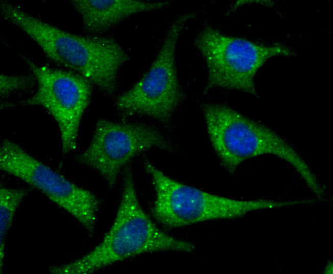
Fig4: ICC staining Apolipoprotein A I in SH-SY-5Y cells (green). The nuclear counter stain is DAPI (blue). Cells were fixed in paraformaldehyde, permeabilised with 0.25% Triton X100/PBS.Application
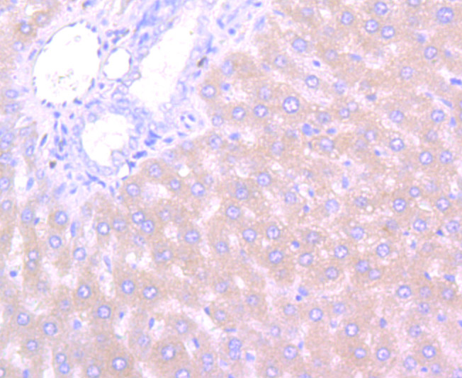
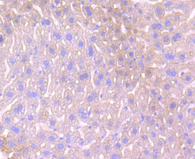
Fig5: Immunohistochemical analysis of paraffin-embedded rat liver tissue using anti-Apolipoprotein A1 antibody. Counter stained with hematoxylin.Application
Fig6: Immunohistochemical analysis of paraffin-embedded rat liver tissue using anti-Apolipoprotein A1 antibody. Counter stained with hematoxylin.Application
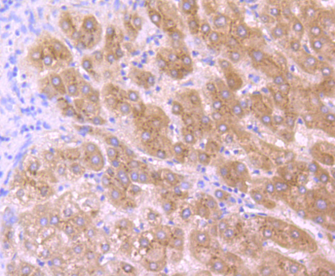
Fig7: Immunohistochemical analysis of paraffin-embedded human liver tissue using anti-Apolipoprotein A I antibody. Counter stained with hematoxylin.Application
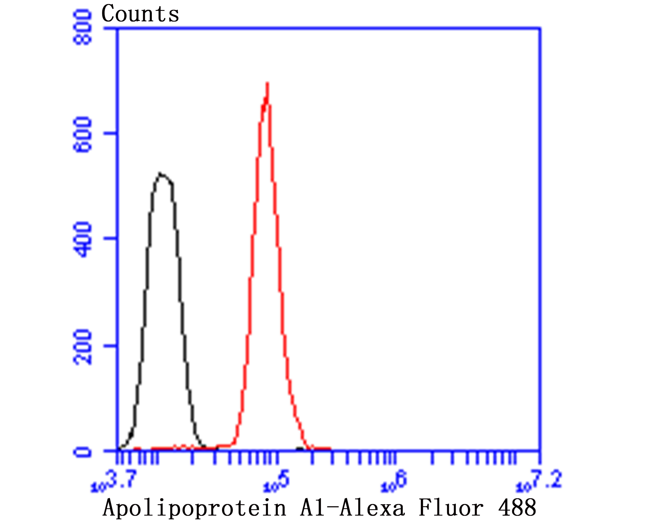
Fig8: Flow cytometric analysis of HepG2 cells with Apolipoprotein A1 antibody at 1/50 dilution (red) compared with an unlabelled control (cells without incubation with primary antibody; black). Alexa Fluor 488-conjugated goat anti-rabbit IgG was used as the secondary antibody.| Product Name | Apolipoprotein A1 |
|---|---|
| Antibody Type | Primary Antibodies |
| Antigen Alias | Apo-AI antibody ApoA I antibody ApoA-I antibody APOA1 antibody APOA1_HUMAN antibody Apolipoprotein A-I(1-242) antibody Apolipoprotein A1 antibody Apolipoprotein AI antibody Brp14 antibody Ltw1 antibody Lvtw1 antibody Sep1 antibody Sep2 antibody |
| Product description | Apolipoproteins are protein components of plasma lipoproteins. The human apoA-I gene encodes a single chain, 243 amino acid protein which promotes cholesterol efflux from tissues to the liver for excretion. Apolipoprotein A-I is the major protein component of high density lipoprotein (HDL) in the plasma. It can function as a cofactor for lecithin cholesterolacyltransferase (LCAT), which is responsible for the formation of most plasma cholesteryl esters. The human apoA-II gene encodes the second most abundant protein of HDL particles, where it influences plasma levels of free fatty acids (FFA). The human apoA-IV gene encodes a 396 amino acid preprotein, which after proteolytic processing is secreted from the intestine in association with chylomicron particles. ApoA-IV is a potent activator of LCAT in vitro. The human apoA-V gene encodes a 366 amino acid protein that is believed to be an important determinant of plasma triglyceride levels. |
| Immunogen | Peptide |
| Clonality | Polyclonal |
|---|---|
| Isotype | IgG |
| Host Species | Rabbit |
| Tested Applications | WBICCIHCFC |
| WB:1:1,000-1:2,000 ICC:1:50-1:200 IHC:1:50-1:100 FC:1:50-1:100 |
|
| Species Reactivity | HumanMouseRat |
| Concentration | 1mg/ml |
| Alternative Names | Apo-AI antibody ApoA I antibody ApoA-I antibody APOA1 antibody APOA1_HUMAN antibody Apolipoprotein A-I(1-242) antibody Apolipoprotein A1 antibody Apolipoprotein AI antibody Brp14 antibody Ltw1 antibody Lvtw1 antibody Sep1 antibody Sep2 antibody |
|---|---|
| Molecular Weight(MW) | 27 kDa |
| Cellular Localization | Secreted. |
Application
Fig1: Western blot analysis of Apolipoprotein A1 on human liver tissue lysate using anti-Apolipoprotein A1 antibody at 1/1,000 dilution.
Application
Fig2: ICC staining Apolipoprotein A1 in HepG2 cells (green). The nuclear counter stain is DAPI (blue). Cells were fixed in paraformaldehyde, permeabilised with 0.25% Triton X100/PBS.
Application
Fig3: ICC staining Apolipoprotein A1 in LOVO cells (green). The nuclear counter stain is DAPI (blue). Cells were fixed in paraformaldehyde, permeabilised with 0.25% Triton X100/PBS.
Application
Fig4: ICC staining Apolipoprotein A I in SH-SY-5Y cells (green). The nuclear counter stain is DAPI (blue). Cells were fixed in paraformaldehyde, permeabilised with 0.25% Triton X100/PBS.
Application
Fig5: Immunohistochemical analysis of paraffin-embedded rat liver tissue using anti-Apolipoprotein A1 antibody. Counter stained with hematoxylin.
Application
Fig6: Immunohistochemical analysis of paraffin-embedded rat liver tissue using anti-Apolipoprotein A1 antibody. Counter stained with hematoxylin.
Application
Fig7: Immunohistochemical analysis of paraffin-embedded human liver tissue using anti-Apolipoprotein A I antibody. Counter stained with hematoxylin.
Application
Fig8: Flow cytometric analysis of HepG2 cells with Apolipoprotein A1 antibody at 1/50 dilution (red) compared with an unlabelled control (cells without incubation with primary antibody; black). Alexa Fluor 488-conjugated goat anti-rabbit IgG was used as the secondary antibody.| Positive Control | Human liver tissue, HepG2, LOVO, SH-SY-5Y, rat liver tissue, mouse liver tissue. |
|---|---|
| Application Notes | WB:1:1,000-1:2,000 ICC:1:50-1:200 IHC:1:50-1:100 FC:1:50-1:100 |
| Form | Liquid |
|---|---|
| Storage Instructions | Store at +4℃ after thawing. Aliquot store at -20℃ or -80℃. Avoid repeated freeze / thaw cycles. |
| Storage Buffer | 1*TBS (pH7.4), 0.5%BSA, 50%Glycerol. Preservative: 0.05% Sodium Azide. |


