Application
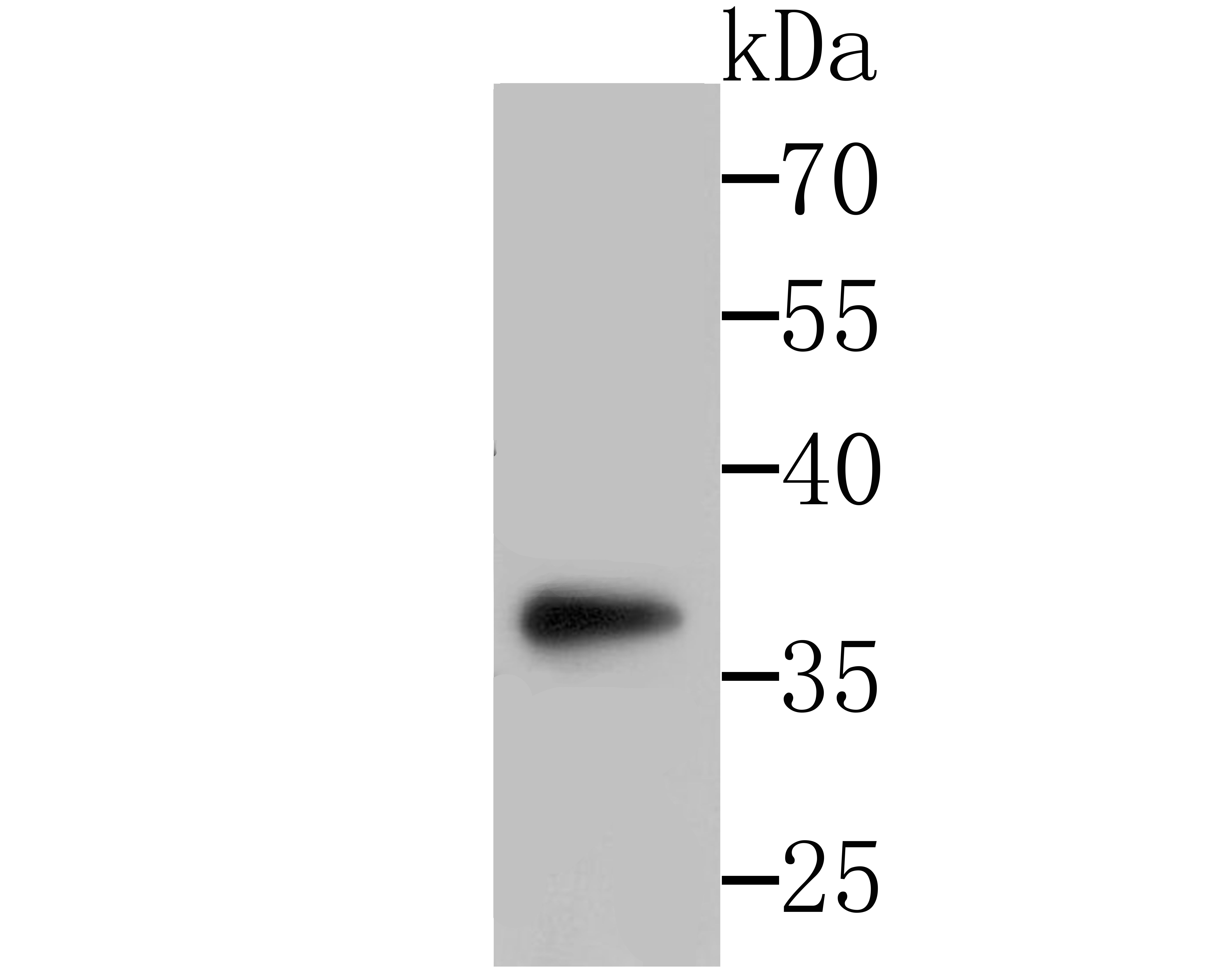
Fig1: Western blot analysis of SFRP1 on SH-SY-5Y cells lysates using anti-SFRP1 antibody at 1/500 dilution.Application
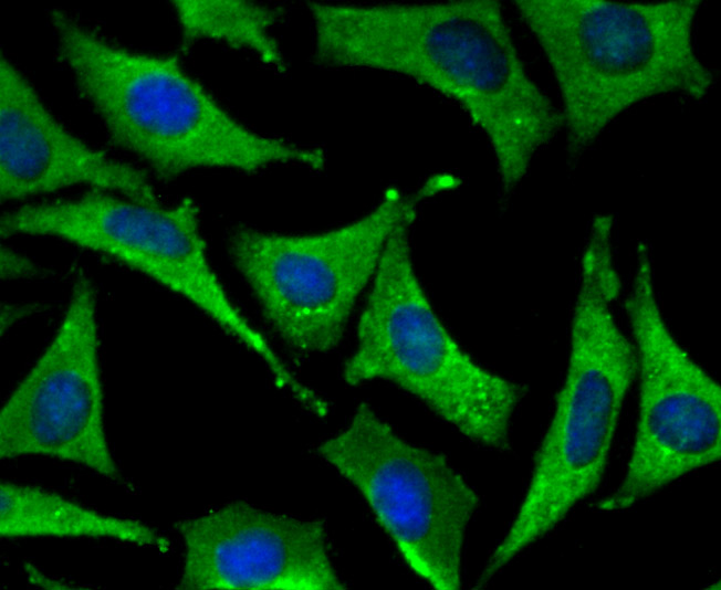
Fig2: ICC staining SFRP1 in SH-SY-5Y cells (green). The nuclear counter stain is DAPI (blue). Cells were fixed in paraformaldehyde, permeabilised with 0.25% Triton X100/PBS.Application
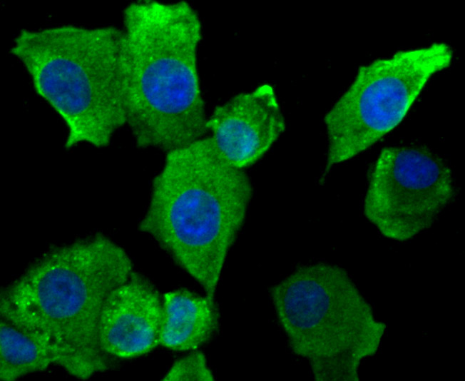
Fig3: ICC staining SFRP1 in A549 cells (green). The nuclear counter stain is DAPI (blue). Cells were fixed in paraformaldehyde, permeabilised with 0.25% Triton X100/PBS.Application
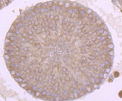
Fig4: Immunohistochemical analysis of paraffin-embedded rat testis tissue using anti-SFRP1 antibody. Counter stained with hematoxylin.Application
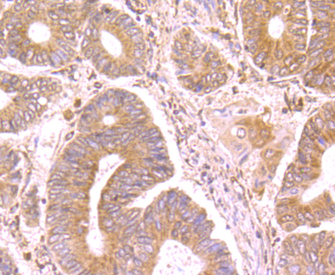
Fig5: Immunohistochemical analysis of paraffin-embedded human colon cancer tissue using anti-SFRP1 antibody. Counter stained with hematoxylin.Application
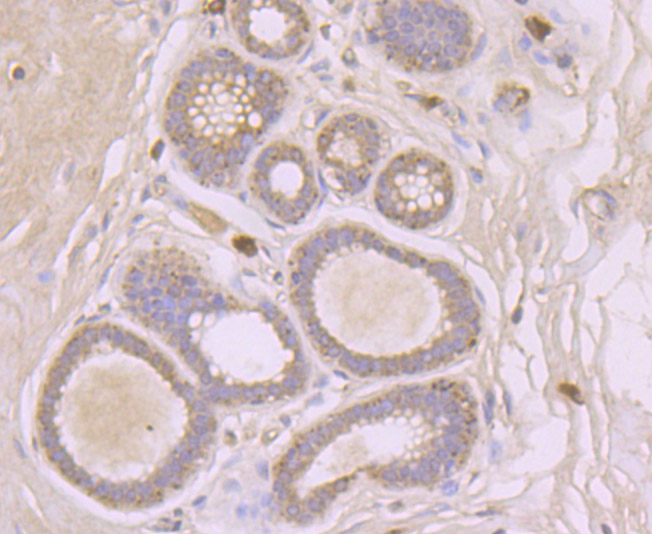
Fig6: Immunohistochemical analysis of paraffin-embedded human breast tissue using anti-SFRP1 antibody. Counter stained with hematoxylin.Application
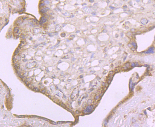
Fig7: Immunohistochemical analysis of paraffin-embedded human placenta tissue using anti-SFRP1 antibody. Counter stained with hematoxylin.Application
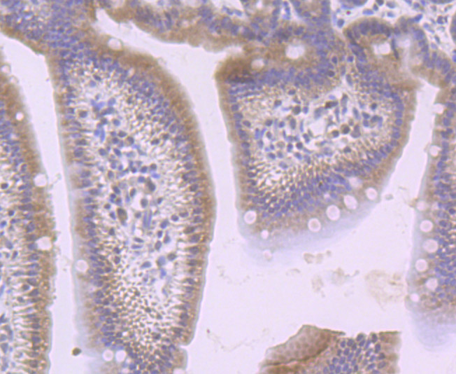
Fig8: Immunohistochemical analysis of paraffin-embedded mouse small intestine tissue using anti-SFRP1 antibody. Counter stained with hematoxylin.Application
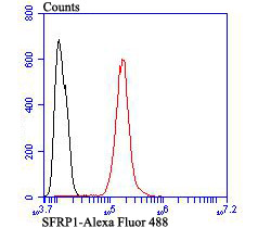
Fig9: Flow cytometric analysis of SH-SY-5Y cells with SFRP1 antibody at 1/100 dilution (red) compared with an unlabelled control (cells without incubation with primary antibody; black). Alexa Fluor 488-conjugated goat anti-rabbit IgG was used as the secondary antibody.| Product Name | SFRP1 Rabbit Polyclonal Antibody |
|---|---|
| Antibody Type | Primary Antibodies |
| Product description | Soluble frizzled-related proteins (sFRPS) function as modulators of Wnt signaling through direct interaction with Wnts. They have a role in regulating cell growth and differentiation in specific cell types. SFRP1 decreases intracellular beta-catenin levels. Has antiproliferative effects on vascular cells, in vitro and in vivo, and can induce, in vivo, an angiogenic response. In vascular cell cycle, delays the G1 phase and entry into the S phase. In kidney development, inhibits tubule formation and bud growth in metanephroi. Inhibits WNT1/WNT4-mediated TCF-dependent transcription. |
| Immunogen | Synthetic peptide within N-terminal human SFRP1. |
| Clonality | Polyclonal |
|---|---|
| Isotype | IgG |
| Host Species | Rabbit |
| Tested Applications | FCICC/IFIHCWB |
| WB:1:500-1:1,000 ICC/IF:1:100-1:500 IHC:1:50-1:200 FC:1:50-1:100 |
|
| Species Reactivity | HumanMouseRat |
| Concentration | 1mg/ml |
| Purification | Protein A |
| Alternative Names | Frizzled related protein 1 antibody FRP 1 antibody FRP antibody FRP-1 antibody FRP1 antibody FrzA antibody SARP 2 antibody SARP-2 antibody SARP2 antibody Secreted apoptosis related protein 2 antibody Secreted apoptosis-related protein 2 antibody Secreted frizzled related protein 1 antibody Secreted frizzled related protein antibody Secreted frizzled-related protein 1 antibody SFRP 1 antibody sFRP-1 antibody SFRP1 antibody SFRP1_HUMAN antibody |
|---|---|
| Molecular Weight(MW) | 35 kDa |
| Cellular Localization | Secreted. |
Application
Fig1: Western blot analysis of SFRP1 on SH-SY-5Y cells lysates using anti-SFRP1 antibody at 1/500 dilution.
Application
Fig2: ICC staining SFRP1 in SH-SY-5Y cells (green). The nuclear counter stain is DAPI (blue). Cells were fixed in paraformaldehyde, permeabilised with 0.25% Triton X100/PBS.
Application
Fig3: ICC staining SFRP1 in A549 cells (green). The nuclear counter stain is DAPI (blue). Cells were fixed in paraformaldehyde, permeabilised with 0.25% Triton X100/PBS.
Application
Fig4: Immunohistochemical analysis of paraffin-embedded rat testis tissue using anti-SFRP1 antibody. Counter stained with hematoxylin.
Application
Fig5: Immunohistochemical analysis of paraffin-embedded human colon cancer tissue using anti-SFRP1 antibody. Counter stained with hematoxylin.
Application
Fig6: Immunohistochemical analysis of paraffin-embedded human breast tissue using anti-SFRP1 antibody. Counter stained with hematoxylin.
Application
Fig7: Immunohistochemical analysis of paraffin-embedded human placenta tissue using anti-SFRP1 antibody. Counter stained with hematoxylin.
Application
Fig8: Immunohistochemical analysis of paraffin-embedded mouse small intestine tissue using anti-SFRP1 antibody. Counter stained with hematoxylin.
Application
Fig9: Flow cytometric analysis of SH-SY-5Y cells with SFRP1 antibody at 1/100 dilution (red) compared with an unlabelled control (cells without incubation with primary antibody; black). Alexa Fluor 488-conjugated goat anti-rabbit IgG was used as the secondary antibody.| Positive Control | A549, SH-SY-5Y, rat testis tissue, human colon cancer tissue, human breast tissue, human placenta tissue, mouse small intestine tissue. |
|---|---|
| Application Notes | WB:1:500-1:1,000 ICC/IF:1:100-1:500 IHC:1:50-1:200 FC:1:50-1:100 |
| Form | Liquid |
|---|---|
| Storage Instructions | Store at +4℃ after thawing. Aliquot store at -20℃ or -80℃. Avoid repeated freeze / thaw cycles. |
| Storage Buffer | 1*TBS (pH7.4), 0.5%BSA, 50%Glycerol. Preservative: 0.05% Sodium Azide. |


