Application
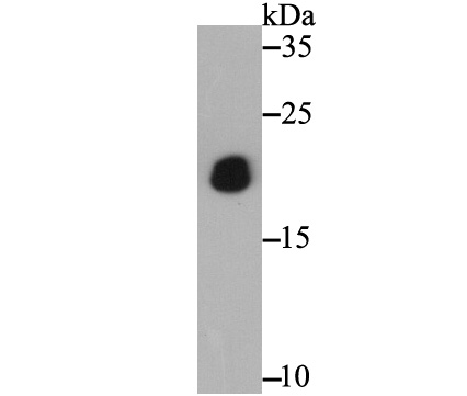
Fig1: Western blot analysis of PTP1B on recombinant protein lysate using anti-PTP1B antibody at 1/5,000 dilution.Application
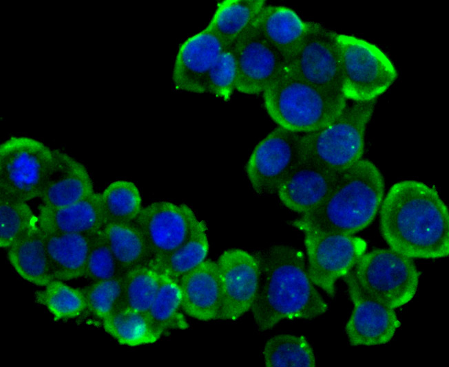
Fig2: ICC staining PTP1B in LOVO cells (green). The nuclear counter stain is DAPI (blue). Cells were fixed in paraformaldehyde, permeabilised with 0.25% Triton X100/PBS.Application
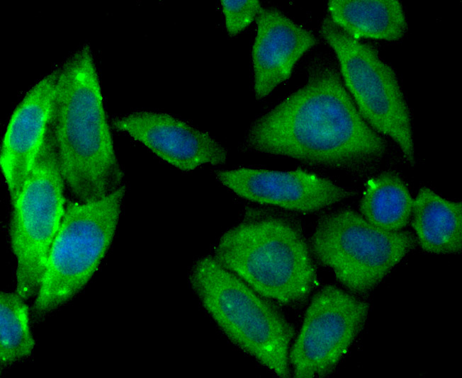
Fig3: ICC staining PTP1B in Siha cells (green). The nuclear counter stain is DAPI (blue). Cells were fixed in paraformaldehyde, permeabilised with 0.25% Triton X100/PBS.Application
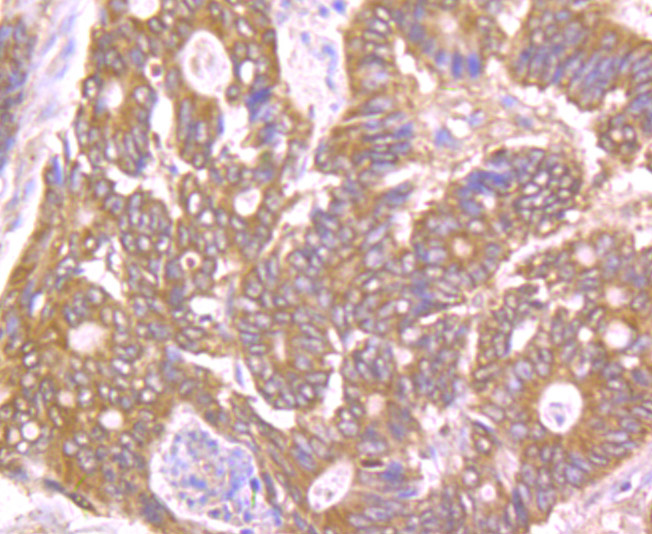
Fig4: Immunohistochemical analysis of paraffin-embedded human colon cancer tissue using anti-PTP1B antibody. Counter stained with hematoxylin.Application
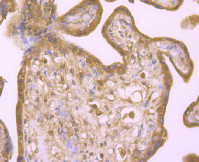
Fig5: Immunohistochemical analysis of paraffin-embedded human placenta tissue using anti-PTP1B antibody. Counter stained with hematoxylin.Application
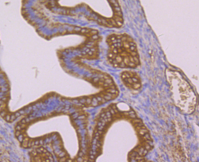
Fig6: Immunohistochemical analysis of paraffin-embedded mouse fallopian tubes tissue using anti-PTP1B antibody. Counter stained with hematoxylin.| Product Name | PTP1B |
|---|---|
| Antibody Type | Primary Antibodies |
| Antigen Alias | PTP1B antibody Non receptor tyrosine phosphatase 1 antibody Protein phosphotyrosylphosphatase 1B antibody Protein tyrosine phosphatase 1B antibody Protein tyrosine phosphatase non receptor type 1 antibody Protein tyrosine phosphatase placental antibody Protein-tyrosine phosphatase 1B antibody PTN1_HUMAN antibody PTP 1B antibody PTP-1B antibody PTPN 1 antibody PTPN1 antibody Tyrosine protein phosphatase non receptor type 1 antibody Tyrosine-protein phosphatase non-receptor type 1 antibody |
| Product description | The phosphorylation of proteins at tyrosine residues has long been recognized as an important regulatory component of signal transduction. This is a reversible process, involving both enzymes that phosphorylate proteins on tyrosine residues as well as a rapidly expanding family of protein tyrosine phosphatases. These latter enzymes bear little resemblance to either the protein serine and protein threonine phosphatases or to the acid and alkaline phosphatases. In most tissues, the major PTPase is a vanadate- and molybdate-sensitive protein. On the basis of sequence analysis, PTP1B (PTPase 1B) expressed in human placenta exhibits similarities both with the common leukocyte antigen (CD45) and with LAR, a homolog of the neural adhesion molecule (NCAM). PTP1B is synthesized as a 435 amino acid precursor protein which is cleaved to generate the active 321 amino acid enzyme. |
| Immunogen | Recombinant protein |
| Clonality | Polyclonal |
|---|---|
| Isotype | IgG |
| Host Species | Rabbit |
| Tested Applications | WBICCIHC |
| WB:1:1,000-1 5,000ICC:1:500-1:2,000 IHC:1:100-1:500 |
|
| Species Reactivity | HumanMouse |
| Concentration | 1mg/ml |
| Alternative Names | PTP1B antibody Non receptor tyrosine phosphatase 1 antibody Protein phosphotyrosylphosphatase 1B antibody Protein tyrosine phosphatase 1B antibody Protein tyrosine phosphatase non receptor type 1 antibody Protein tyrosine phosphatase placental antibody Protein-tyrosine phosphatase 1B antibody PTN1_HUMAN antibody PTP 1B antibody PTP-1B antibody PTPN 1 antibody PTPN1 antibody Tyrosine protein phosphatase non receptor type 1 antibody Tyrosine-protein phosphatase non-receptor type 1 antibody |
|---|---|
| Molecular Weight(MW) | 50 kDa |
| Cellular Localization | Endoplasmic reticulum. |
Application
Fig1: Western blot analysis of PTP1B on recombinant protein lysate using anti-PTP1B antibody at 1/5,000 dilution.
Application
Fig2: ICC staining PTP1B in LOVO cells (green). The nuclear counter stain is DAPI (blue). Cells were fixed in paraformaldehyde, permeabilised with 0.25% Triton X100/PBS.
Application
Fig3: ICC staining PTP1B in Siha cells (green). The nuclear counter stain is DAPI (blue). Cells were fixed in paraformaldehyde, permeabilised with 0.25% Triton X100/PBS.
Application
Fig4: Immunohistochemical analysis of paraffin-embedded human colon cancer tissue using anti-PTP1B antibody. Counter stained with hematoxylin.
Application
Fig5: Immunohistochemical analysis of paraffin-embedded human placenta tissue using anti-PTP1B antibody. Counter stained with hematoxylin.
Application
Fig6: Immunohistochemical analysis of paraffin-embedded mouse fallopian tubes tissue using anti-PTP1B antibody. Counter stained with hematoxylin.| Positive Control | Recombinant protein lysate, LOVO, SiHa, human colon cancer tissue, human placenta tissue, mouse fallopian tubes tissue. |
|---|---|
| Application Notes | WB:1:1,000-1 5,000ICC:1:500-1:2,000 IHC:1:100-1:500 |
| Form | Liquid |
|---|---|
| Storage Instructions | Store at +4℃ after thawing. Aliquot store at -20℃ or -80℃. Avoid repeated freeze / thaw cycles. |
| Storage Buffer | 1*TBS (pH7.4), 0.5%BSA, 50%Glycerol. Preservative: 0.05% Sodium Azide. |


