Application
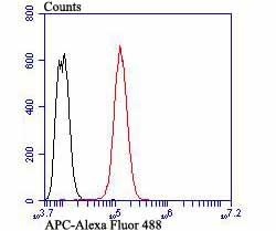
Fig8: Flow cytometric analysis of SH-SY-5Y cells with APC antibody at 1/100 dilution (red) compared with an unlabelled control (cells without incubation with primary antibody; black). Alexa Fluor 488-conjugated goat anti-rabbit IgG was used as the secondary antibody.Application
Fig8: Flow cytometric analysis of SH-SY-5Y cells with APC antibody at 1/100 dilution (red) compared with an unlabelled control (cells without incubation with primary antibody; black). Alexa Fluor 488-conjugated goat anti-rabbit IgG was used as the secondary antibody.Application
Fig8: Flow cytometric analysis of SH-SY-5Y cells with APC antibody at 1/100 dilution (red) compared with an unlabelled control (cells without incubation with primary antibody; black). Alexa Fluor 488-conjugated goat anti-rabbit IgG was used as the secondary antibody.Application
Fig8: Flow cytometric analysis of SH-SY-5Y cells with APC antibody at 1/100 dilution (red) compared with an unlabelled control (cells without incubation with primary antibody; black). Alexa Fluor 488-conjugated goat anti-rabbit IgG was used as the secondary antibody.Application
Fig8: Flow cytometric analysis of SH-SY-5Y cells with APC antibody at 1/100 dilution (red) compared with an unlabelled control (cells without incubation with primary antibody; black). Alexa Fluor 488-conjugated goat anti-rabbit IgG was used as the secondary antibody.Application
Fig8: Flow cytometric analysis of SH-SY-5Y cells with APC antibody at 1/100 dilution (red) compared with an unlabelled control (cells without incubation with primary antibody; black). Alexa Fluor 488-conjugated goat anti-rabbit IgG was used as the secondary antibody.Application
Fig8: Flow cytometric analysis of SH-SY-5Y cells with APC antibody at 1/100 dilution (red) compared with an unlabelled control (cells without incubation with primary antibody; black). Alexa Fluor 488-conjugated goat anti-rabbit IgG was used as the secondary antibody.Application
Fig8: Flow cytometric analysis of SH-SY-5Y cells with APC antibody at 1/100 dilution (red) compared with an unlabelled control (cells without incubation with primary antibody; black). Alexa Fluor 488-conjugated goat anti-rabbit IgG was used as the secondary antibody.| Product Name | APC |
|---|---|
| Antibody Type | Primary Antibodies |
| Antigen Alias | Adenomatous Polyposis Coli antibody Adenomatous polyposis coli protein antibody Apc antibody APC_HUMAN antibody CC1 antibody Deleted in polyposis 2.5 antibody DP2 antibody DP2.5 antibody DP3 antibody FAP antibody FPC antibody GS antibody Protein APC antibody |
| Product description | The adenomatous polyposis syndromes, familial adenomatous polyposis (FAP) and Gardner's syndrome (GS), are characterized by numerous adenomatous polyps throughout the entire colon. These polyps invariably progress to colon cancer in addition to other extracolonic manifestations. The cloning of the APC gene revealed a ubiquitously expressed protein, 2,843 amino acids in length, which is frequently mutated in patients suffering from FAP and GS. APC has been found to be associated with structural components of intracellular junctions. β-catenin and γ-catenin (also called plakoglobin), are involved in the regulation of cellular adhesion. APC and E-cadherin compete for binding to specific internal regions of both β- and γ-catenin. Interactions between cytoskeleton and the APC, E-cadherin, β/γ catenin complex are mediated by α-catenin. |
| Immunogen | Peptide |
| Clonality | Polyclonal |
|---|---|
| Isotype | IgG |
| Host Species | Rabbit |
| Tested Applications | WBICCIHCFC |
| ICC:1:50-1:200 IHC:1:100-1:200 FC:1:50-1:100 WB:1:500 |
|
| Species Reactivity | HumanMouseRat |
| Concentration | 1mg/ml |
| Alternative Names | Adenomatous Polyposis Coli antibody Adenomatous polyposis coli protein antibody Apc antibody APC_HUMAN antibody CC1 antibody Deleted in polyposis 2.5 antibody DP2 antibody DP2.5 antibody DP3 antibody FAP antibody FPC antibody GS antibody Protein APC antibody |
|---|---|
| Molecular Weight(MW) | Predicted band size 312 kDa |
| Cellular Localization | Cell junction. Cytoplasm. Cytoskeleton. Membrane. |
Application
Fig8: Flow cytometric analysis of SH-SY-5Y cells with APC antibody at 1/100 dilution (red) compared with an unlabelled control (cells without incubation with primary antibody; black). Alexa Fluor 488-conjugated goat anti-rabbit IgG was used as the secondary antibody.
Application
Fig8: Flow cytometric analysis of SH-SY-5Y cells with APC antibody at 1/100 dilution (red) compared with an unlabelled control (cells without incubation with primary antibody; black). Alexa Fluor 488-conjugated goat anti-rabbit IgG was used as the secondary antibody.
Application
Fig8: Flow cytometric analysis of SH-SY-5Y cells with APC antibody at 1/100 dilution (red) compared with an unlabelled control (cells without incubation with primary antibody; black). Alexa Fluor 488-conjugated goat anti-rabbit IgG was used as the secondary antibody.
Application
Fig8: Flow cytometric analysis of SH-SY-5Y cells with APC antibody at 1/100 dilution (red) compared with an unlabelled control (cells without incubation with primary antibody; black). Alexa Fluor 488-conjugated goat anti-rabbit IgG was used as the secondary antibody.
Application
Fig8: Flow cytometric analysis of SH-SY-5Y cells with APC antibody at 1/100 dilution (red) compared with an unlabelled control (cells without incubation with primary antibody; black). Alexa Fluor 488-conjugated goat anti-rabbit IgG was used as the secondary antibody.
Application
Fig8: Flow cytometric analysis of SH-SY-5Y cells with APC antibody at 1/100 dilution (red) compared with an unlabelled control (cells without incubation with primary antibody; black). Alexa Fluor 488-conjugated goat anti-rabbit IgG was used as the secondary antibody.
Application
Fig8: Flow cytometric analysis of SH-SY-5Y cells with APC antibody at 1/100 dilution (red) compared with an unlabelled control (cells without incubation with primary antibody; black). Alexa Fluor 488-conjugated goat anti-rabbit IgG was used as the secondary antibody.
Application
Fig8: Flow cytometric analysis of SH-SY-5Y cells with APC antibody at 1/100 dilution (red) compared with an unlabelled control (cells without incubation with primary antibody; black). Alexa Fluor 488-conjugated goat anti-rabbit IgG was used as the secondary antibody.| Positive Control | HUVEC, SH-SY-5Y, SiHa, rat brain tissue, human kidney tissue, human breast tissue, mouse colon tissue. |
|---|---|
| Application Notes | ICC:1:50-1:200 IHC:1:100-1:200 FC:1:50-1:100 WB:1:500 |
| Form | Liquid |
|---|---|
| Storage Instructions | Store at +4℃ after thawing. Aliquot store at -20℃ or -80℃. Avoid repeated freeze / thaw cycles. |
| Storage Buffer | 1*TBS (pH7.4), 0.5%BSA, 50%Glycerol. Preservative: 0.05% Sodium Azide. |


