Application
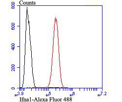
Fig8: Flow cytometric analysis of SH-SY5Y cells with IFNA1 antibody at 1/100 dilution (red) compared with an unlabelled control (cells without incubation with primary antibody; black). Alexa Fluor 488-conjugated goat anti rabbit IgG was used as the secondary antibody.Application
Fig8: Flow cytometric analysis of SH-SY5Y cells with IFNA1 antibody at 1/100 dilution (red) compared with an unlabelled control (cells without incubation with primary antibody; black). Alexa Fluor 488-conjugated goat anti rabbit IgG was used as the secondary antibody.Application
Fig8: Flow cytometric analysis of SH-SY5Y cells with IFNA1 antibody at 1/100 dilution (red) compared with an unlabelled control (cells without incubation with primary antibody; black). Alexa Fluor 488-conjugated goat anti rabbit IgG was used as the secondary antibody.Application
Fig8: Flow cytometric analysis of SH-SY5Y cells with IFNA1 antibody at 1/100 dilution (red) compared with an unlabelled control (cells without incubation with primary antibody; black). Alexa Fluor 488-conjugated goat anti rabbit IgG was used as the secondary antibody.Application
Fig8: Flow cytometric analysis of SH-SY5Y cells with IFNA1 antibody at 1/100 dilution (red) compared with an unlabelled control (cells without incubation with primary antibody; black). Alexa Fluor 488-conjugated goat anti rabbit IgG was used as the secondary antibody.Application
Fig8: Flow cytometric analysis of SH-SY5Y cells with IFNA1 antibody at 1/100 dilution (red) compared with an unlabelled control (cells without incubation with primary antibody; black). Alexa Fluor 488-conjugated goat anti rabbit IgG was used as the secondary antibody.Application
Fig8: Flow cytometric analysis of SH-SY5Y cells with IFNA1 antibody at 1/100 dilution (red) compared with an unlabelled control (cells without incubation with primary antibody; black). Alexa Fluor 488-conjugated goat anti rabbit IgG was used as the secondary antibody.Application
Fig8: Flow cytometric analysis of SH-SY5Y cells with IFNA1 antibody at 1/100 dilution (red) compared with an unlabelled control (cells without incubation with primary antibody; black). Alexa Fluor 488-conjugated goat anti rabbit IgG was used as the secondary antibody.| Product Name | Ifna1 |
|---|---|
| Antibody Type | Primary Antibodies |
| Antigen Alias | IFN-alpha-14 antibody IFN14_HUMAN antibody IFNA14 antibody Interferon alpha 14 antibody Interferon alpha H antibody Interferon alpha-14 antibody Interferon alpha-H antibody Interferon lambda-2-H antibody Interferon lambda2H antibody LeIF H antibody LeIFH antibody MGC125756 antibody MGC125757 antibody |
| Product description | The genes encoding type I interferons (IFNs), which include 14 IFN-α genes (two of which are IFN-α1 and IFN-α13), one IFN-β gene, one IFN-w (also known as IFN-α II1) gene and a number of IFN-w pseudogenes, are clustered on human chromosome 9. IFN-α and -β are cytokines that are widely known to induce potent antiviral activity. They exert a variety of other biological effects, including antitumor and immunomodulatory activities and are increasingly used clinically to treat a range of malignancies, myelodysplasias and autoimmune diseases. IFN-w is antigenically different from human IFN-α, IFN-β or IFN-γ, but is a component of natural mixtures of IFN species produced by virus-induced leukocytes or Burkitt's lymphoma cells. The type I interferon receptor (IFN-αR) interacts with IFN-α, IFN-β and IFN-w, and seems to be a multisubunit receptor. |
| Immunogen | Peptide |
| Clonality | Polyclonal |
|---|---|
| Isotype | IgG |
| Host Species | Rabbit |
| Tested Applications | WBICCIHCFC |
| WB:1:500 ICC:1:50-1:200 IHC:1:50-1:200 FC:1:50-1:100 |
|
| Species Reactivity | HumanMouseRat |
| Concentration | 1mg/ml |
| Alternative Names | IFN-alpha-14 antibody IFN14_HUMAN antibody IFNA14 antibody Interferon alpha 14 antibody Interferon alpha H antibody Interferon alpha-14 antibody Interferon alpha-H antibody Interferon lambda-2-H antibody Interferon lambda2H antibody LeIF H antibody LeIFH antibody MGC125756 antibody MGC125757 antibody |
|---|---|
| Molecular Weight(MW) | 22 kDa |
| Cellular Localization | Secreted. |
Application
Fig8: Flow cytometric analysis of SH-SY5Y cells with IFNA1 antibody at 1/100 dilution (red) compared with an unlabelled control (cells without incubation with primary antibody; black). Alexa Fluor 488-conjugated goat anti rabbit IgG was used as the secondary antibody.
Application
Fig8: Flow cytometric analysis of SH-SY5Y cells with IFNA1 antibody at 1/100 dilution (red) compared with an unlabelled control (cells without incubation with primary antibody; black). Alexa Fluor 488-conjugated goat anti rabbit IgG was used as the secondary antibody.
Application
Fig8: Flow cytometric analysis of SH-SY5Y cells with IFNA1 antibody at 1/100 dilution (red) compared with an unlabelled control (cells without incubation with primary antibody; black). Alexa Fluor 488-conjugated goat anti rabbit IgG was used as the secondary antibody.
Application
Fig8: Flow cytometric analysis of SH-SY5Y cells with IFNA1 antibody at 1/100 dilution (red) compared with an unlabelled control (cells without incubation with primary antibody; black). Alexa Fluor 488-conjugated goat anti rabbit IgG was used as the secondary antibody.
Application
Fig8: Flow cytometric analysis of SH-SY5Y cells with IFNA1 antibody at 1/100 dilution (red) compared with an unlabelled control (cells without incubation with primary antibody; black). Alexa Fluor 488-conjugated goat anti rabbit IgG was used as the secondary antibody.
Application
Fig8: Flow cytometric analysis of SH-SY5Y cells with IFNA1 antibody at 1/100 dilution (red) compared with an unlabelled control (cells without incubation with primary antibody; black). Alexa Fluor 488-conjugated goat anti rabbit IgG was used as the secondary antibody.
Application
Fig8: Flow cytometric analysis of SH-SY5Y cells with IFNA1 antibody at 1/100 dilution (red) compared with an unlabelled control (cells without incubation with primary antibody; black). Alexa Fluor 488-conjugated goat anti rabbit IgG was used as the secondary antibody.
Application
Fig8: Flow cytometric analysis of SH-SY5Y cells with IFNA1 antibody at 1/100 dilution (red) compared with an unlabelled control (cells without incubation with primary antibody; black). Alexa Fluor 488-conjugated goat anti rabbit IgG was used as the secondary antibody.| Positive Control | Mouse liver tissue lysate, N2A, SHG-44, SH-SY5Y, rat brain tissue, human kidney tissue, mouse brain tissue. |
|---|---|
| Application Notes | WB:1:500 ICC:1:50-1:200 IHC:1:50-1:200 FC:1:50-1:100 |
| Form | Liquid |
|---|---|
| Storage Instructions | Store at +4℃ after thawing. Aliquot store at -20℃ or -80℃. Avoid repeated freeze / thaw cycles. |
| Storage Buffer | 1*TBS (pH7.4), 0.5%BSA, 50%Glycerol. Preservative: 0.05% Sodium Azide. |


