Application
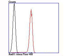
Fig8: Flow cytometric analysis of Jurkat cells with Rad51 antibody at 1/100 dilution (red) compared with an unlabelled control (cells without incubation with primary antibody; black).Application
Fig8: Flow cytometric analysis of Jurkat cells with Rad51 antibody at 1/100 dilution (red) compared with an unlabelled control (cells without incubation with primary antibody; black).Application
Fig8: Flow cytometric analysis of Jurkat cells with Rad51 antibody at 1/100 dilution (red) compared with an unlabelled control (cells without incubation with primary antibody; black).Application
Fig8: Flow cytometric analysis of Jurkat cells with Rad51 antibody at 1/100 dilution (red) compared with an unlabelled control (cells without incubation with primary antibody; black).Application
Fig8: Flow cytometric analysis of Jurkat cells with Rad51 antibody at 1/100 dilution (red) compared with an unlabelled control (cells without incubation with primary antibody; black).Application
Fig8: Flow cytometric analysis of Jurkat cells with Rad51 antibody at 1/100 dilution (red) compared with an unlabelled control (cells without incubation with primary antibody; black).Application
Fig8: Flow cytometric analysis of Jurkat cells with Rad51 antibody at 1/100 dilution (red) compared with an unlabelled control (cells without incubation with primary antibody; black).Application
Fig8: Flow cytometric analysis of Jurkat cells with Rad51 antibody at 1/100 dilution (red) compared with an unlabelled control (cells without incubation with primary antibody; black).| Product Name | Rad51 [JM54-26] |
|---|---|
| Antibody Type | Primary Antibodies |
| Antigen Alias | BRCA1/BRCA2 containing complex, subunit 5 antibody BRCC 5 antibody BRCC5 antibody DNA repair protein RAD51 homolog 1 antibody DNA repair protein rhp51 antibody FANCR antibody hRAD51 antibody HsRAD51 antibody HsT16930 antibody MRMV2 antibody Rad 51 antibody RAD51 antibody RAD51 homolog (RecA homolog, E. coli) (S. cerevisiae) antibody RAD51 homolog A antibody RAD51 homolog antibody RAD51 recombinase antibody RAD51, S. cerevisiae, homolog of antibody RAD51_HUMAN antibody RAD51A antibody RECA antibody RecA like protein antibody RecA, E. coli, homolog of antibody Recombination protein A antibody |
| Product description | Rad51 (RECA, BRCC5) interacts with BRCA1 and BRCA2 to influence subcellular localization and cellular response to DNA damage. BRCA2 inactivation may be a key event leading to genomic instability and tumorigenesis from deregulation of Rad51. Rad52 forms a heptameric ring that binds single-stranded DNA ends and catalyzes DNA-DNA interaction necessary for the annealing of complementary strands. Rad52 can interact with Rad51. Rad54A of the DEAD-like helicase superfamily binds to double-strand DNA and induces a DNA topological change, which is thought to facilitate homologous DNA pairing and stimulate DNA recombination. Rad54B of the DEAD-like helicase superfamily binds to double-stranded DNA and displays ATPase activity in the presence of DNA. Rad54B is abundant in testis and spleen, and mutations of this gene occur in primary lymphoma and colon cancer. MRE11 (meiotic recombination 11, ATLD, HNGS1) is a nuclear 3′-5′ exonuclease/endonuclease that associates with Rad50 and influences homologous recombination, telomere length maintenance, and DNA double-strand break repair. MRE11 is most abundant in proliferating tissues. |
| Immunogen | Recombinant protein |
| Clonality | Monoclonal |
|---|---|
| Isotype | IgG |
| Host Species | Recombinant rabbit |
| Tested Applications | WBICC/IFIHCIPFC |
| WB:1:500 ICC/IF:1:50-1:200 IHC:1:50-1:200 IP:1:10-1:50 FC:1:50-1:100 |
|
| Species Reactivity | HumanMouseRat |
| Concentration | 1mg/ml |
| Alternative Names | BRCA1/BRCA2 containing complex subunit 5 antibody BRCC 5 antibody BRCC5 antibody DNA repair protein RAD51 homolog 1 antibody DNA repair protein rhp51 antibody FANCR antibody hRAD51 antibody HsRAD51 antibody HsT16930 antibody MRMV2 antibody Rad 51 antibody RAD51 antibody RAD51 homolog (RecA homolog E. coli) (S. cerevisiae) antibody RAD51 homolog A antibody RAD51 homolog antibody RAD51 recombinase antibody RAD51 S. cerevisiae homolog of antibody RAD51_HUMAN antibody RAD51A antibody RECA antibody RecA like protein antibody RecA E. coli homolog of antibody Recombination protein A antibody |
|---|---|
| Molecular Weight(MW) | 37 kDa |
| Cellular Localization | Nucleus. Cytoplasm. |
Application
Fig8: Flow cytometric analysis of Jurkat cells with Rad51 antibody at 1/100 dilution (red) compared with an unlabelled control (cells without incubation with primary antibody; black).
Application
Fig8: Flow cytometric analysis of Jurkat cells with Rad51 antibody at 1/100 dilution (red) compared with an unlabelled control (cells without incubation with primary antibody; black).
Application
Fig8: Flow cytometric analysis of Jurkat cells with Rad51 antibody at 1/100 dilution (red) compared with an unlabelled control (cells without incubation with primary antibody; black).
Application
Fig8: Flow cytometric analysis of Jurkat cells with Rad51 antibody at 1/100 dilution (red) compared with an unlabelled control (cells without incubation with primary antibody; black).
Application
Fig8: Flow cytometric analysis of Jurkat cells with Rad51 antibody at 1/100 dilution (red) compared with an unlabelled control (cells without incubation with primary antibody; black).
Application
Fig8: Flow cytometric analysis of Jurkat cells with Rad51 antibody at 1/100 dilution (red) compared with an unlabelled control (cells without incubation with primary antibody; black).
Application
Fig8: Flow cytometric analysis of Jurkat cells with Rad51 antibody at 1/100 dilution (red) compared with an unlabelled control (cells without incubation with primary antibody; black).
Application
Fig8: Flow cytometric analysis of Jurkat cells with Rad51 antibody at 1/100 dilution (red) compared with an unlabelled control (cells without incubation with primary antibody; black).| Positive Control | Mouse testie tissue lytase, LOVO, PANC-1, rat epididymis tissue, human placenta tissue, mouse testis tissue, Jurkat. |
|---|---|
| Application Notes | WB:1:500 ICC/IF:1:50-1:200 IHC:1:50-1:200 IP:1:10-1:50 FC:1:50-1:100 |
| Form | Liquid |
|---|---|
| Storage Instructions | Store at +4℃ after thawing. Aliquot store at -20℃ or -80℃. Avoid repeated freeze / thaw cycles. |
| Storage Buffer | 1*TBS (pH7.4), 1%BSA, 40%Glycerol. Preservative: 0.05% Sodium Azide. |


