Application
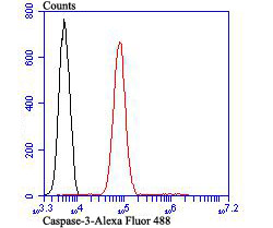
Fig6: Flow cytometric analysis of Jurkat cells with Caspase-3 antibody at 1/100 dilution (red) compared with an unlabelled control (cells without incubation with primary antibody; black). Alexa Fluor 488-conjugated goat anti rabbit IgG was used as the secondary antibody.Application
Fig6: Flow cytometric analysis of Jurkat cells with Caspase-3 antibody at 1/100 dilution (red) compared with an unlabelled control (cells without incubation with primary antibody; black). Alexa Fluor 488-conjugated goat anti rabbit IgG was used as the secondary antibody.Application
Fig6: Flow cytometric analysis of Jurkat cells with Caspase-3 antibody at 1/100 dilution (red) compared with an unlabelled control (cells without incubation with primary antibody; black). Alexa Fluor 488-conjugated goat anti rabbit IgG was used as the secondary antibody.Application
Fig6: Flow cytometric analysis of Jurkat cells with Caspase-3 antibody at 1/100 dilution (red) compared with an unlabelled control (cells without incubation with primary antibody; black). Alexa Fluor 488-conjugated goat anti rabbit IgG was used as the secondary antibody.Application
Fig6: Flow cytometric analysis of Jurkat cells with Caspase-3 antibody at 1/100 dilution (red) compared with an unlabelled control (cells without incubation with primary antibody; black). Alexa Fluor 488-conjugated goat anti rabbit IgG was used as the secondary antibody.Application
Fig6: Flow cytometric analysis of Jurkat cells with Caspase-3 antibody at 1/100 dilution (red) compared with an unlabelled control (cells without incubation with primary antibody; black). Alexa Fluor 488-conjugated goat anti rabbit IgG was used as the secondary antibody.| Product Name | Caspase-3 |
|---|---|
| Antibody Type | Primary Antibodies |
| Antigen Alias | A830040C14Rik antibody Apopain antibody CASP-3 antibody CASP3 antibody CASP3_HUMAN antibody Casp3a antibody Caspase 3 antibody Caspase 3, apoptosis-related cysteine peptidase antibody Caspase 3, apoptosis-related cysteine protease antibody Caspase 3, apoptosis-related cysteine protease a antibody Caspase-3 subunit p12 antibody CC3 antibody CPP-32 antibody CPP32 antibody CPP32B antibody Cysteine protease CPP32 antibody EC 3.4.22.56 antibody LICE antibody mldy antibody OTTHUMP00000165052 antibody OTTHUMP00000165053 antibody OTTHUMP00000165054 antibody PARP cleavage protease antibody Procaspase3 antibody Protein Yama antibody SCA 1 antibody SCA-1 antibody SREBP cleavage activity 1 antibody Yama antibody |
| Product description | Involved in the activation cascade of caspases responsible for apoptosis execution. At the onset of apoptosis it proteolytically cleaves poly(ADP-ribose) polymerase (PARP) at a '216-Asp-|-Gly-217' bond. Cleaves and activates sterol regulatory element binding proteins (SREBPs) between the basic helix-loop-helix leucine zipper domain and the membrane attachment domain. Cleaves and activates caspase-6, -7 and -9. Involved in the cleavage of huntingtin. Triggers cell adhesion in sympathetic neurons through RET cleavage. |
| Immunogen | Peptide |
| Clonality | Polyclonal |
|---|---|
| Isotype | IgG |
| Host Species | Rabbit |
| Tested Applications | WBICCIHCFC |
| WB:1:500-1:2,000 ICC:1:50-1:200 IHC:1:50-1:200 FC:1:50-1:100 |
|
| Species Reactivity | HumanMouse |
| Concentration | 1mg/ml |
| Alternative Names | A830040C14Rik antibody
Apopain antibody
CASP-3 antibody
CASP3 antibody
CASP3_HUMAN antibody
Casp3a antibody
Caspase 3 antibody
Caspase 3 apoptosis-related cysteine peptidase antibody Caspase 3 apoptosis-related cysteine protease antibody Caspase 3 apoptosis-related cysteine protease a antibody Caspase-3 subunit p12 antibody CC3 antibody CPP-32 antibody CPP32 antibody CPP32B antibody Cysteine protease CPP32 antibody EC 3.4.22.56 antibody LICE antibody mldy antibody OTTHUMP00000165052 antibody OTTHUMP00000165053 antibody OTTHUMP00000165054 antibody PARP cleavage protease antibody Procaspase3 antibody Protein Yama antibody SCA 1 antibody SCA-1 antibody SREBP cleavage activity 1 antibody Yama antibody |
|---|---|
| Molecular Weight(MW) | 32 kDa |
| Cellular Localization | Cytoplasm. |
Application
Fig6: Flow cytometric analysis of Jurkat cells with Caspase-3 antibody at 1/100 dilution (red) compared with an unlabelled control (cells without incubation with primary antibody; black). Alexa Fluor 488-conjugated goat anti rabbit IgG was used as the secondary antibody.
Application
Fig6: Flow cytometric analysis of Jurkat cells with Caspase-3 antibody at 1/100 dilution (red) compared with an unlabelled control (cells without incubation with primary antibody; black). Alexa Fluor 488-conjugated goat anti rabbit IgG was used as the secondary antibody.
Application
Fig6: Flow cytometric analysis of Jurkat cells with Caspase-3 antibody at 1/100 dilution (red) compared with an unlabelled control (cells without incubation with primary antibody; black). Alexa Fluor 488-conjugated goat anti rabbit IgG was used as the secondary antibody.
Application
Fig6: Flow cytometric analysis of Jurkat cells with Caspase-3 antibody at 1/100 dilution (red) compared with an unlabelled control (cells without incubation with primary antibody; black). Alexa Fluor 488-conjugated goat anti rabbit IgG was used as the secondary antibody.
Application
Fig6: Flow cytometric analysis of Jurkat cells with Caspase-3 antibody at 1/100 dilution (red) compared with an unlabelled control (cells without incubation with primary antibody; black). Alexa Fluor 488-conjugated goat anti rabbit IgG was used as the secondary antibody.
Application
Fig6: Flow cytometric analysis of Jurkat cells with Caspase-3 antibody at 1/100 dilution (red) compared with an unlabelled control (cells without incubation with primary antibody; black). Alexa Fluor 488-conjugated goat anti rabbit IgG was used as the secondary antibody.| Positive Control | A549, HepG2, HUVEC, Jurkat, human tonsil tissue, human spleen tissue. |
|---|---|
| Application Notes | WB:1:500-1:2,000 ICC:1:50-1:200 IHC:1:50-1:200 FC:1:50-1:100 |
| Form | Liquid |
|---|---|
| Storage Instructions | Store at +4℃ after thawing. Aliquot store at -20℃ or -80℃. Avoid repeated freeze / thaw cycles. |
| Storage Buffer | 1*TBS (pH7.4), 0.5%BSA, 50%Glycerol. Preservative: 0.05% Sodium Azide. |


