Application
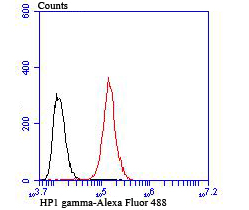
Fig6: Flow cytometric analysis of Hela cells with HP1 gamma antibody at 1/100 dilution (red) compared with an unlabelled control (cells without incubation with primary antibody; black). Alexa Fluor® 488-conjugated goat anti-rabbit IgG was used as the secondary antibody.Application
Fig6: Flow cytometric analysis of Hela cells with HP1 gamma antibody at 1/100 dilution (red) compared with an unlabelled control (cells without incubation with primary antibody; black). Alexa Fluor® 488-conjugated goat anti-rabbit IgG was used as the secondary antibody.Application
Fig6: Flow cytometric analysis of Hela cells with HP1 gamma antibody at 1/100 dilution (red) compared with an unlabelled control (cells without incubation with primary antibody; black). Alexa Fluor® 488-conjugated goat anti-rabbit IgG was used as the secondary antibody.Application
Fig6: Flow cytometric analysis of Hela cells with HP1 gamma antibody at 1/100 dilution (red) compared with an unlabelled control (cells without incubation with primary antibody; black). Alexa Fluor® 488-conjugated goat anti-rabbit IgG was used as the secondary antibody.Application
Fig6: Flow cytometric analysis of Hela cells with HP1 gamma antibody at 1/100 dilution (red) compared with an unlabelled control (cells without incubation with primary antibody; black). Alexa Fluor® 488-conjugated goat anti-rabbit IgG was used as the secondary antibody.Application
Fig6: Flow cytometric analysis of Hela cells with HP1 gamma antibody at 1/100 dilution (red) compared with an unlabelled control (cells without incubation with primary antibody; black). Alexa Fluor® 488-conjugated goat anti-rabbit IgG was used as the secondary antibody.| Product Name | HP1 gamma [JU81-36] |
|---|---|
| Antibody Type | Primary Antibodies |
| Antigen Alias | CBX 3 antibody CBX3 antibody CBX3_HUMAN antibody Chromobox homolog 3 (HP1 gamma homolog, Drosophila) antibody Chromobox homolog 3 antibody Chromobox protein homolog 3 antibody GAMMA antibody HECH antibody Heterochromatin like protein 1 antibody Heterochromatin protein 1 homolog gamma antibody Heterochromatin protein HP1 gamma antibody HP1 gamma antibody HP1 gamma homolog antibody HP1Hs gamma antibody Modifier 2 protein antibody |
| Product description | inaccessible to DNA-modifying reagents. In late S phase, p150 directly associates with heterochromatin associated proteins 1 (HP1α, HP1β and HP1γ). As cells prepare for mitosis, CAF-1 p150 and some HP1 progressively dissociate from heterochromatin, coinciding with the phosphorylation of Histone H3. The HP1 proteins reassociate with chromatin at the end of mitosis, as Histone H3 is dephosphorylated. |
| Immunogen | Recombinant protein |
| Clonality | Monoclonal |
|---|---|
| Isotype | IgG |
| Host Species | Recombinant rabbit |
| Tested Applications | WBIHCFCIP |
| WB:1:500 IHC:1:50-1:200 FC:1:50-1:100 IP:1:10-1:50 |
|
| Species Reactivity | HumanMouseRat |
| Concentration | 1mg/ml |
| Alternative Names | CBX 3 antibody
CBX3 antibody
CBX3_HUMAN antibody
Chromobox homolog 3 (HP1 gamma homolog Drosophila) antibody Chromobox homolog 3 antibody Chromobox protein homolog 3 antibody GAMMA antibody HECH antibody Heterochromatin like protein 1 antibody Heterochromatin protein 1 homolog gamma antibody Heterochromatin protein HP1 gamma antibody HP1 gamma antibody HP1 gamma homolog antibody HP1Hs gamma antibody Modifier 2 protein antibody |
|---|---|
| Molecular Weight(MW) | 20 kDa |
| Cellular Localization | Nucleus. |
Application
Fig6: Flow cytometric analysis of Hela cells with HP1 gamma antibody at 1/100 dilution (red) compared with an unlabelled control (cells without incubation with primary antibody; black). Alexa Fluor® 488-conjugated goat anti-rabbit IgG was used as the secondary antibody.
Application
Fig6: Flow cytometric analysis of Hela cells with HP1 gamma antibody at 1/100 dilution (red) compared with an unlabelled control (cells without incubation with primary antibody; black). Alexa Fluor® 488-conjugated goat anti-rabbit IgG was used as the secondary antibody.
Application
Fig6: Flow cytometric analysis of Hela cells with HP1 gamma antibody at 1/100 dilution (red) compared with an unlabelled control (cells without incubation with primary antibody; black). Alexa Fluor® 488-conjugated goat anti-rabbit IgG was used as the secondary antibody.
Application
Fig6: Flow cytometric analysis of Hela cells with HP1 gamma antibody at 1/100 dilution (red) compared with an unlabelled control (cells without incubation with primary antibody; black). Alexa Fluor® 488-conjugated goat anti-rabbit IgG was used as the secondary antibody.
Application
Fig6: Flow cytometric analysis of Hela cells with HP1 gamma antibody at 1/100 dilution (red) compared with an unlabelled control (cells without incubation with primary antibody; black). Alexa Fluor® 488-conjugated goat anti-rabbit IgG was used as the secondary antibody.
Application
Fig6: Flow cytometric analysis of Hela cells with HP1 gamma antibody at 1/100 dilution (red) compared with an unlabelled control (cells without incubation with primary antibody; black). Alexa Fluor® 488-conjugated goat anti-rabbit IgG was used as the secondary antibody.| Positive Control | PC-12, MCF-7, rat testis tissue, human tonsil tissue, human placenta tissue, mouse colon tissue, Hela. |
|---|---|
| Application Notes | WB:1:500 IHC:1:50-1:200 FC:1:50-1:100 IP:1:10-1:50 |
| Form | Liquid |
|---|---|
| Storage Instructions | Store at +4℃ after thawing. Aliquot store at -20℃ or -80℃. Avoid repeated freeze / thaw cycles. |
| Storage Buffer | 1*TBS (pH7.4), 1%BSA, 40%Glycerol. Preservative: 0.05% Sodium Azide. |


