Application
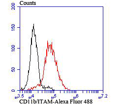
Fig4: Flow cytometric analysis of THP-1 cells with CD11b antibody at 1/100 dilution (red) compared with an unlabelled control (cells without incubation with primary antibody; black). Alexa Fluor 488-conjugated goat anti rabbit IgG was used as the secondary antibody.Application
Fig4: Flow cytometric analysis of THP-1 cells with CD11b antibody at 1/100 dilution (red) compared with an unlabelled control (cells without incubation with primary antibody; black). Alexa Fluor 488-conjugated goat anti rabbit IgG was used as the secondary antibody.Application
Fig4: Flow cytometric analysis of THP-1 cells with CD11b antibody at 1/100 dilution (red) compared with an unlabelled control (cells without incubation with primary antibody; black). Alexa Fluor 488-conjugated goat anti rabbit IgG was used as the secondary antibody.Application
Fig4: Flow cytometric analysis of THP-1 cells with CD11b antibody at 1/100 dilution (red) compared with an unlabelled control (cells without incubation with primary antibody; black). Alexa Fluor 488-conjugated goat anti rabbit IgG was used as the secondary antibody.| Product Name | CD11b [JU93-81] |
|---|---|
| Antibody Type | Primary Antibodies |
| Antigen Alias | antigen CD11b (p170) antibody Antigen CD11b p170 antibody CD11 antigen like family member B antibody CD11 antigen-like family member B antibody CD11b antibody CD11b/CD18 antibody CD49d antibody Cell surface glycoprotein MAC-1 subunit alpha antibody Complement component 3 receptor 3 subunit antibody Complement Component Receptor 3 Alpha antibody Complement receptor type 3, alpha subunit antibody CR 3 alpha chain (CR3A) antibody CR 3 alpha chain antibody CR-3 alpha chain antibody CR3 antibody CR3A antibody F730045J24Rik antibody Integrin Alpha M antibody Integrin alpha M chain antibody Integrin alpha-M antibody Integrin beta 2 alpha subunit antibody Integrin subunit alpha M antibody integrin, alpha M (complement component 3 receptor 3 subunit) antibody ITAM_HUMAN antibody ITGAM antibody Leukocyte adhesion receptor MO1 antibody Ly-40 antibody MAC 1 antibody Mac-1a antibody MAC1 antibody Mac1, alpha subunit antibody MAC1A antibody Macrophage antigen alpha polypeptide antibody MGC117044 antibody Mo1, alpha subunit antibody MO1A antibody Neutrophil adherence receptor alpha M subunit antibody Neutrophil adherence receptor antibody SLEB6 antibody |
| Product description | Integrin αM, also designated complement component receptor-3 α, CD11b (p170), macrophage antigen a polypeptide, cell surface glycoprotein Mac-1 a subunit, MAC1A, MO1A and ITGAM) is a cell adhesion molecule that acts as a receptor for cell surface ligands such as intracellular adhesion molecules (ICAMs) or soluble ligands. Integrins are heterodimeric proteins that contain an a chain and b chain. Integrin αM combines with the Integrin β2 to form a leukocyte-specific integrin referred to as macrophage receptor 1 (Mac-1), or inactivated-C3b (iC3b) receptor 3 (CR3). Integrin αM/β2 is important in the adherence of neutrophils and monocytes to stimulated endothelium, and also in the phagocytosis of complement coated particles. |
| Immunogen | Recombinant protein |
| Clonality | Monoclonal |
|---|---|
| Isotype | IgG |
| Host Species | Recombinant rabbit |
| Tested Applications | WBICCIFIPIHCFC |
| ICC:1:50-1:200 IHC:1:50-1:200 FC:1:50-1:100 WB:1:500-1:1,000 |
|
| Species Reactivity | Human |
| Concentration | 1mg/ml |
| Alternative Names | antigen CD11b (p170) antibody
Antigen CD11b p170 antibody
CD11 antigen like family member B antibody
CD11 antigen-like family member B antibody
CD11b antibody
CD11b/CD18 antibody
CD49d antibody
Cell surface glycoprotein MAC-1 subunit alpha antibody
Complement component 3 receptor 3 subunit antibody
Complement Component Receptor 3 Alpha antibody
Complement receptor type 3 alpha subunit antibody CR 3 alpha chain (CR3A) antibody CR 3 alpha chain antibody CR-3 alpha chain antibody CR3 antibody CR3A antibody F730045J24Rik antibody Integrin Alpha M antibody Integrin alpha M chain antibody Integrin alpha-M antibody Integrin beta 2 alpha subunit antibody Integrin subunit alpha M antibody integrin alpha M (complement component 3 receptor 3 subunit) antibody ITAM_HUMAN antibody ITGAM antibody Leukocyte adhesion receptor MO1 antibody Ly-40 antibody MAC 1 antibody Mac-1a antibody MAC1 antibody Mac1 alpha subunit antibody MAC1A antibody Macrophage antigen alpha polypeptide antibody MGC117044 antibody Mo1 alpha subunit antibody MO1A antibody Neutrophil adherence receptor alpha M subunit antibody Neutrophil adherence receptor antibody SLEB6 antibody |
|---|---|
| Molecular Weight(MW) | 170 kDa |
| Cellular Localization | Membrane. |
| SwissProt ID | P11215 |
|---|
Application
Fig4: Flow cytometric analysis of THP-1 cells with CD11b antibody at 1/100 dilution (red) compared with an unlabelled control (cells without incubation with primary antibody; black). Alexa Fluor 488-conjugated goat anti rabbit IgG was used as the secondary antibody.
Application
Fig4: Flow cytometric analysis of THP-1 cells with CD11b antibody at 1/100 dilution (red) compared with an unlabelled control (cells without incubation with primary antibody; black). Alexa Fluor 488-conjugated goat anti rabbit IgG was used as the secondary antibody.
Application
Fig4: Flow cytometric analysis of THP-1 cells with CD11b antibody at 1/100 dilution (red) compared with an unlabelled control (cells without incubation with primary antibody; black). Alexa Fluor 488-conjugated goat anti rabbit IgG was used as the secondary antibody.
Application
Fig4: Flow cytometric analysis of THP-1 cells with CD11b antibody at 1/100 dilution (red) compared with an unlabelled control (cells without incubation with primary antibody; black). Alexa Fluor 488-conjugated goat anti rabbit IgG was used as the secondary antibody.| Positive Control | LOVO, human tonsil tissue, human spleen tissue, THP-1. |
|---|---|
| Application Notes | ICC:1:50-1:200 IHC:1:50-1:200 FC:1:50-1:100 WB:1:500-1:1,000 |
| Form | Liquid |
|---|---|
| Storage Instructions | Store at +4℃ after thawing. Aliquot store at -20℃ or -80℃. Avoid repeated freeze / thaw cycles. |
| Storage Buffer | 1*TBS (pH7.4), 1%BSA, 40%Glycerol. Preservative: 0.05% Sodium Azide. |


