
Application
Fig1: Western blot analysis of CD55 on human plasma using anti-CD55 antibody at 1/1,000Application
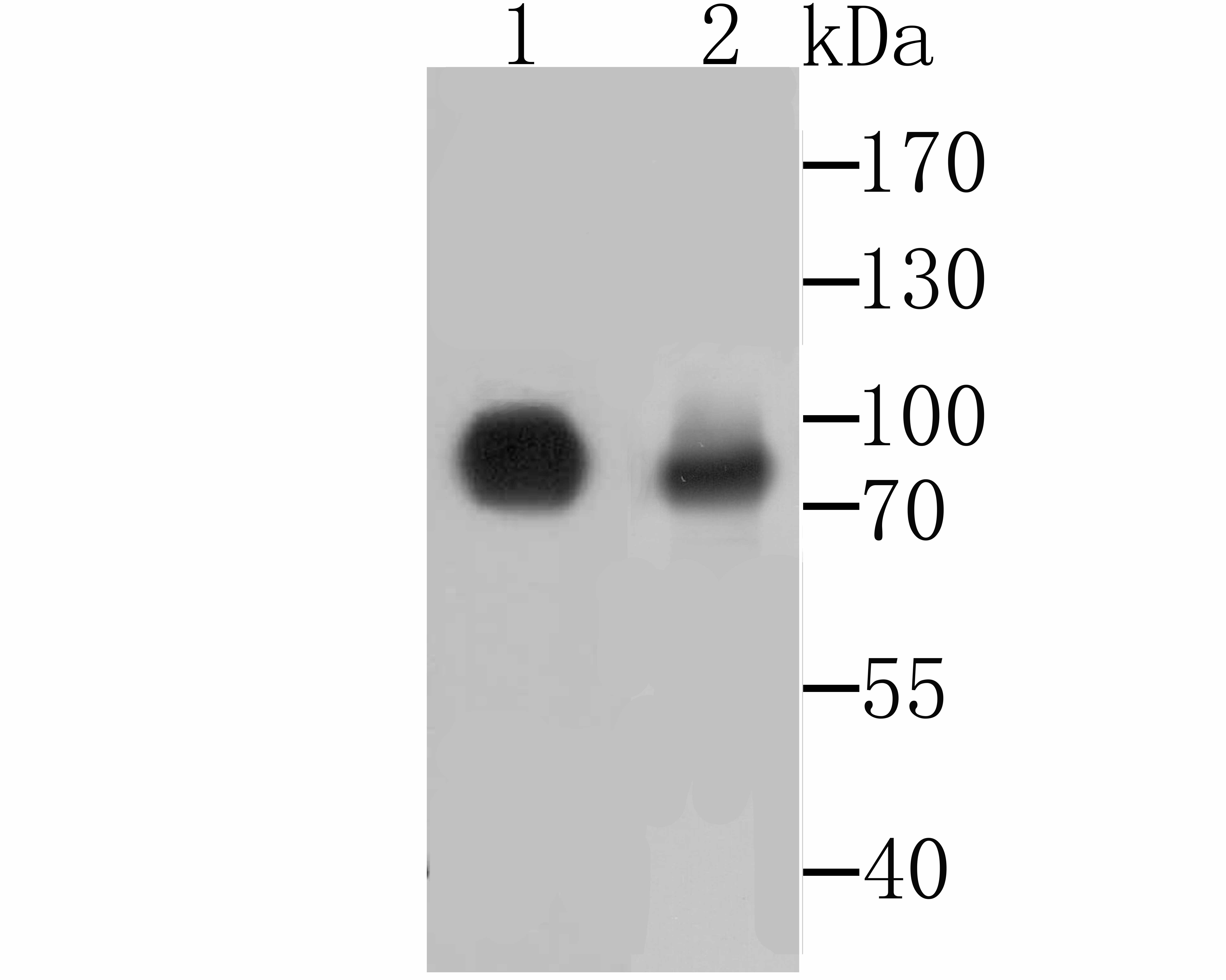
Fig2: Western blot analysis of CD55 on Hela and PC-3M cell lysates using anti-CD55 antibody at 1/500 dilution.Application
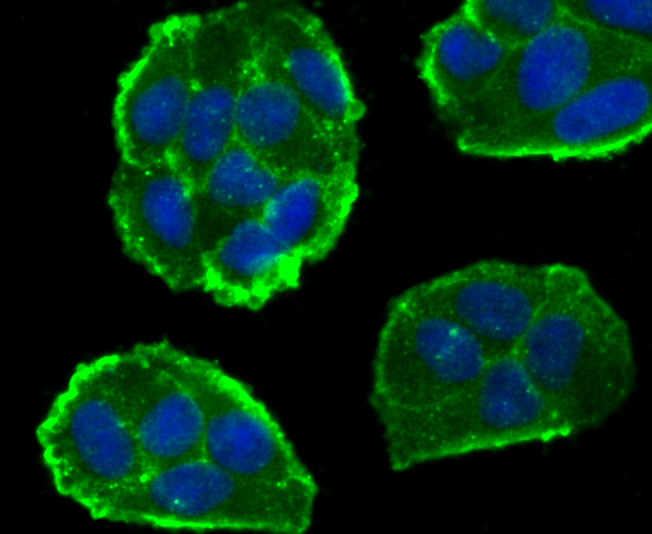
Fig3: ICC staining CD55 in Hela cells (green). The nuclear counter stain is DAPI (blue). Cells were fixed in paraformaldehyde, permeabilised with 0.25% Triton X100/PBS.Application
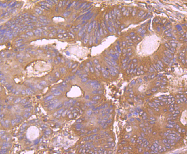
Fig4: Immunohistochemical analysis of paraffin-embedded human colon cancer tissue using anti-CD55 antibody. Counter stained with hematoxylin.Application
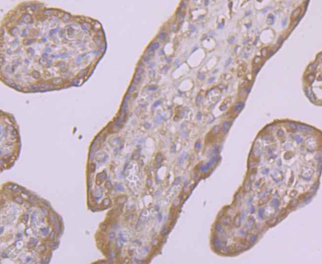
Fig5: Immunohistochemical analysis of paraffin-embedded human placenta tissue using anti-CD55 antibody. Counter stained with hematoxylin.| Product Name | CD55 Rabbit Polyclonal Antibody |
|---|---|
| Antibody Type | Primary Antibodies |
| Product description | CD55, also called decay accelerating factor (DAF), is a GPI-anchored single chain glycoprotein. CD55 may play a role in protecting cells from complement-mediated lysis by preventing the amplification steps of the complement cascade. CD55 functions to prevent the assembly of C3 convertase or to accelerate the disassembly of preformed convertase, which blocks formation of the membrane attack complex. CD55 is expressed on cells in contact with serum, including hematopoietic and many non-hematopoietic cells. |
| Immunogen | Recombinant protein with Human CD55 aa 150-380. |
| Clonality | Polyclonal |
|---|---|
| Isotype | IgG |
| Host Species | Rabbit |
| Tested Applications | ICCIHCWB |
| WB:1:500-1:1,000 ICC:1:50-1:200 IHC:1:50-1:200 |
|
| Species Reactivity | Human |
| Concentration | 1mg/ml |
| Alternative Names | CD 55 antibody
CD55 antibody
CD55 antigen antibody
CD55 Cromer blood group system antibody
CD55 molecule (Cromer blood group) antibody
CD55 molecule antibody
CD55 molecule decay accelerating factor for complement (Cromer blood group) antibody Cd55a antibody Complement decay accelerating factor antibody Complement decay-accelerating factor antibody Complement decay-accelerating factor GPI-anchored antibody CR antibody CROM antibody Cromer Blood Group antigen antibody Cromer blood group system antibody DAF antibody Daf-GPI antibody DAF_HUMAN antibody Daf1 antibody Dcay accelerating factor for complement (CD55 Cromer blood group system) antibody Decay accelarating factor 1 isoform CRA_a antibody Decay accelerating factor (GPI-form) antibody Decay Accelerating Factor for Complement antibody Decay accelerating factor GPI-form antibody Decay accelerating factor soluble-form antibody GPI-DAF antibody TC antibody |
|---|---|
| Molecular Weight(MW) | 42/49/60 kDa (Predicted band size) |
| Cellular Localization | Cell membrane. Secreted. |
| SwissProt ID | P08174 |
|---|
Application
Fig1: Western blot analysis of CD55 on human plasma using anti-CD55 antibody at 1/1,000
Application
Fig2: Western blot analysis of CD55 on Hela and PC-3M cell lysates using anti-CD55 antibody at 1/500 dilution.
Application
Fig3: ICC staining CD55 in Hela cells (green). The nuclear counter stain is DAPI (blue). Cells were fixed in paraformaldehyde, permeabilised with 0.25% Triton X100/PBS.
Application
Fig4: Immunohistochemical analysis of paraffin-embedded human colon cancer tissue using anti-CD55 antibody. Counter stained with hematoxylin.
Application
Fig5: Immunohistochemical analysis of paraffin-embedded human placenta tissue using anti-CD55 antibody. Counter stained with hematoxylin.| Positive Control | Hela and PC-3M cell lysates, human plasma, human colon cancer tissue, human placenta tissue. |
|---|---|
| Application Notes | WB:1:500-1:1,000 ICC:1:50-1:200 IHC:1:50-1:200 |
| Form | Liquid |
|---|---|
| Storage Instructions | Store at +4℃ after thawing. Aliquot store at -20℃. Avoid repeated freeze / thaw cycles. |
| Storage Buffer | 1*TBS (pH7.4), 0.5%BSA, 50%Glycerol. Preservative: 0.05% Sodium Azide. |


