Application
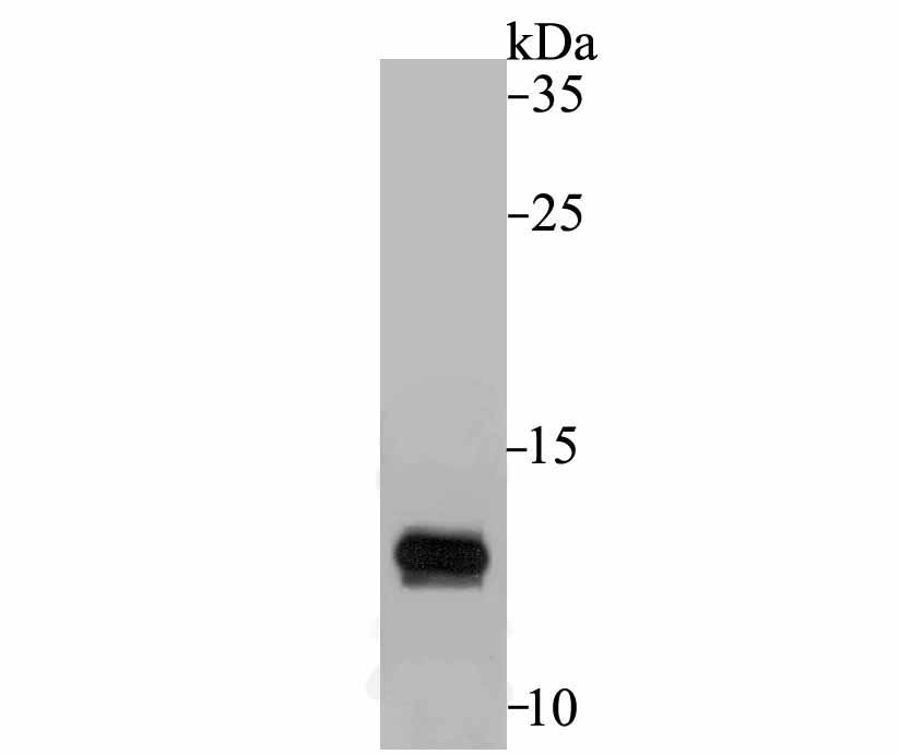
Fig1: Western blot analysis of APOC3 on human liver tissue lysate using anti-APOC3 antibody at 1/500 dilution.Application
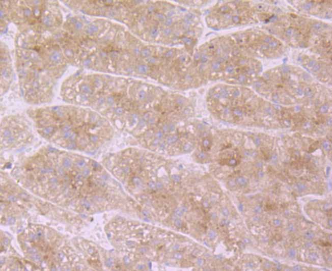
Fig2: Immunohistochemical analysis of paraffin-embedded human liver tissue using anti-APOC3 antibody. Counter stained with hematoxylin.Application
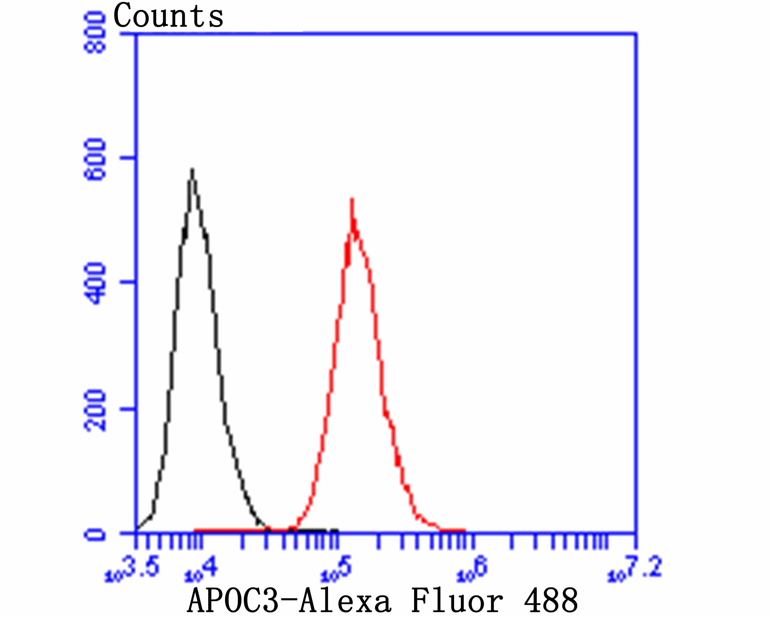
Fig3: Flow cytometric analysis of HepG2 cells with APOC3 antibody at 1/100 dilution (red) compared with an unlabelled control (cells without incubation with primary antibody; black). Alexa Fluor 488-conjugated goat anti-rabbit IgG was used as the secondary antibody.| Product Name | APOC3 |
|---|---|
| Antibody Type | Primary Antibodies |
| Antigen Alias | APOC3 antibody APO C3 antibody Apo CIII antibody Apo-CIII antibody APOC 3 antibody ApoC III antibody ApoC-III antibody APOC3 antibody APOC3_HUMAN antibody ApoCIII antibody Apolipoprotein C III antibody Apolipoprotein C-III antibody Apolipoprotein C3 antibody ApolipoproteinCIII antibody MGC150353 antibody |
| Product description | Component of triglyceride-rich very low density lipoproteins (VLDL) and high density lipoproteins (HDL) in plasma. Plays a multifaceted role in triglyceride homeostasis. Intracellularly, promotes hepatic very low density lipoprotein 1 (VLDL1) assembly and secretion; extracellularly, attenuates hydrolysis and clearance of triglyceride-rich lipoproteins (TRLs). Impairs the lipolysis of TRLs by inhibiting lipoprotein lipase and the hepatic uptake of TRLs by remnant receptors. Formed of several curved helices connected via semiflexible hinges, so that it can wrap tightly around the curved micelle surface and easily adapt to the different diameters of its natural binding partners. |
| Immunogen | Recombinant protein with Human APOC3 aa 1-100. |
| Clonality | Polyclonal |
|---|---|
| Isotype | IgG |
| Host Species | Rabbit |
| Tested Applications | WBIHCFC |
| WB:1:500-1:2000 IHC:1:50-1:200 FC:1:50-1:100 |
|
| Species Reactivity | Human |
| Concentration | 1mg/ml |
| Alternative Names | APOC3 antibody APO C3 antibody Apo CIII antibody Apo-CIII antibody APOC 3 antibody ApoC III antibody ApoC-III antibody APOC3 antibody APOC3_HUMAN antibody ApoCIII antibody Apolipoprotein C III antibody Apolipoprotein C-III antibody Apolipoprotein C3 antibody ApolipoproteinCIII antibody MGC150353 antibody |
|---|---|
| Molecular Weight(MW) | 11 kDa |
| Cellular Localization | Secreted. |
| SwissProt ID | P02656 |
|---|
Application
Fig1: Western blot analysis of APOC3 on human liver tissue lysate using anti-APOC3 antibody at 1/500 dilution.
Application
Fig2: Immunohistochemical analysis of paraffin-embedded human liver tissue using anti-APOC3 antibody. Counter stained with hematoxylin.
Application
Fig3: Flow cytometric analysis of HepG2 cells with APOC3 antibody at 1/100 dilution (red) compared with an unlabelled control (cells without incubation with primary antibody; black). Alexa Fluor 488-conjugated goat anti-rabbit IgG was used as the secondary antibody.| Positive Control | Human liver tissue, HepG2. |
|---|---|
| Application Notes | WB:1:500-1:2000 IHC:1:50-1:200 FC:1:50-1:100 |
| Form | Liquid |
|---|---|
| Storage Instructions | Store at +4℃ after thawing. Aliquot store at -20℃. Avoid repeated freeze / thaw cycles. |
| Storage Buffer | 1*TBS (pH7.4), 0.5%BSA, 50%Glycerol. Preservative: 0.05% Sodium Azide. |


