Application
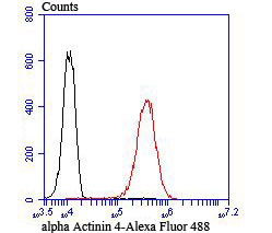
Fig7: Flow cytometric analysis of A549 cells with alpha Actinin 4 antibody at 1/100 dilution (red) compared with an unlabelled control (cells without incubation with primary antibody; black).Application
Fig7: Flow cytometric analysis of A549 cells with alpha Actinin 4 antibody at 1/100 dilution (red) compared with an unlabelled control (cells without incubation with primary antibody; black).Application
Fig7: Flow cytometric analysis of A549 cells with alpha Actinin 4 antibody at 1/100 dilution (red) compared with an unlabelled control (cells without incubation with primary antibody; black).Application
Fig7: Flow cytometric analysis of A549 cells with alpha Actinin 4 antibody at 1/100 dilution (red) compared with an unlabelled control (cells without incubation with primary antibody; black).Application
Fig7: Flow cytometric analysis of A549 cells with alpha Actinin 4 antibody at 1/100 dilution (red) compared with an unlabelled control (cells without incubation with primary antibody; black).Application
Fig7: Flow cytometric analysis of A549 cells with alpha Actinin 4 antibody at 1/100 dilution (red) compared with an unlabelled control (cells without incubation with primary antibody; black).Application
Fig7: Flow cytometric analysis of A549 cells with alpha Actinin 4 antibody at 1/100 dilution (red) compared with an unlabelled control (cells without incubation with primary antibody; black).| Product Name | alpha Actinin 4 [JU20-23] |
|---|---|
| Antibody Type | Primary Antibodies |
| Antigen Alias | actinin 4 antibody Actinin alpha 4 antibody actinin4 antibody ACTN 4 antibody ACTN4 antibody ACTN4_HUMAN antibody Alpha-actinin-4 antibody DKFZp686K23158 antibody F actin cross linking protein antibody F-actin cross-linking protein antibody Focal segmental glomerulosclerosis 1 antibody FSGS 1 antibody FSGS antibody FSGS1 antibody Non muscle alpha actinin 4 antibody Non-muscle alpha-actinin 4 antibody |
| Product description | The spectrin gene family encodes a diverse group of cytoskeletal proteins that include spectrins, dystrophins and α-actinins. There are four tissue-specific α-actinins, namely α-actinin-1, α-actinin-2, α-actinin-3 and α-actinin-4, which are localized to muscle and non-muscle cells, including skeletal, cardiac and smooth muscle cells, as well as within the cytoskeleton. Each α-actinin protein contains one Actin-binding domain, two calponin-homology domains, two EF-hand domains and four spectrin repeats, through which they function as bundling proteins that can cross-link F-Actin, thus anchoring Actin to a variety of intracellular structures. Defects in the gene encoding α-actinin-4 are the cause of focal segmental glomerulosclerosis 1 (FSGS1), a common renal lesion characterized by decreasing kidney function and, ultimately, renal failure. are actually sensitive to the Profilin proteins in these foods. |
| Immunogen | Recombinant protein |
| Clonality | Monoclonal |
|---|---|
| Isotype | IgG |
| Host Species | Recombinant rabbit |
| Tested Applications | WBICCIFIPIHCFC |
| WB:1:500-1:2,000 ICC:1:100-1:500 IHC:1:50-1:200 FC:1:50-1:100 IP:1:10-1:50 |
|
| Species Reactivity | HumanMouseRat |
| Concentration | 1mg/ml |
| Alternative Names | actinin 4 antibody Actinin alpha 4 antibody actinin4 antibody ACTN 4 antibody ACTN4 antibody ACTN4_HUMAN antibody Alpha-actinin-4 antibody DKFZp686K23158 antibody F actin cross linking protein antibody F-actin cross-linking protein antibody Focal segmental glomerulosclerosis 1 antibody FSGS 1 antibody FSGS antibody FSGS1 antibody Non muscle alpha actinin 4 antibody Non-muscle alpha-actinin 4 antibody |
|---|---|
| Molecular Weight(MW) | 105 kDa |
| Cellular Localization | Cell junction. Cytoplasm. Nucleus. |
Application
Fig7: Flow cytometric analysis of A549 cells with alpha Actinin 4 antibody at 1/100 dilution (red) compared with an unlabelled control (cells without incubation with primary antibody; black).
Application
Fig7: Flow cytometric analysis of A549 cells with alpha Actinin 4 antibody at 1/100 dilution (red) compared with an unlabelled control (cells without incubation with primary antibody; black).
Application
Fig7: Flow cytometric analysis of A549 cells with alpha Actinin 4 antibody at 1/100 dilution (red) compared with an unlabelled control (cells without incubation with primary antibody; black).
Application
Fig7: Flow cytometric analysis of A549 cells with alpha Actinin 4 antibody at 1/100 dilution (red) compared with an unlabelled control (cells without incubation with primary antibody; black).
Application
Fig7: Flow cytometric analysis of A549 cells with alpha Actinin 4 antibody at 1/100 dilution (red) compared with an unlabelled control (cells without incubation with primary antibody; black).
Application
Fig7: Flow cytometric analysis of A549 cells with alpha Actinin 4 antibody at 1/100 dilution (red) compared with an unlabelled control (cells without incubation with primary antibody; black).
Application
Fig7: Flow cytometric analysis of A549 cells with alpha Actinin 4 antibody at 1/100 dilution (red) compared with an unlabelled control (cells without incubation with primary antibody; black).| Positive Control | Hela, PC-12, NIH-3T3, rar liver tissue lysate, A431, HepG2, A549, LOVO, human colon cancer tissue, mouse colon tissue. |
|---|---|
| Application Notes | WB:1:500-1:2,000 ICC:1:100-1:500 IHC:1:50-1:200 FC:1:50-1:100 IP:1:10-1:50 |
| Form | Liquid |
|---|---|
| Storage Instructions | Store at +4℃ after thawing. Aliquot store at -20℃ or -80℃. Avoid repeated freeze / thaw cycles. |
| Storage Buffer | 1*TBS (pH7.4), 1%BSA, 40%Glycerol. Preservative: 0.05% Sodium Azide. |


