Application
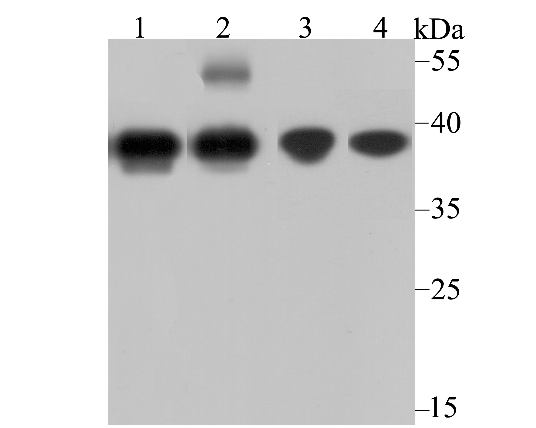
Fig1: Western blot analysis of APE1 on different lysates using anti-APE1 antibody at 1/1,000 dilution. Positive control: Lane1: HL-60 Lane2: Human skin tissue Lane3: MCF-7 Lane4: Mouse placenta tissueApplication
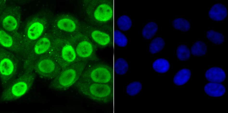
Fig2: ICC staining APE1 in HepG2 cells (green). The nuclear counter stain is DAPI (blue). Cells were fixed in paraformaldehyde, permeabilised with 0.25% Triton X100/PBS.Application
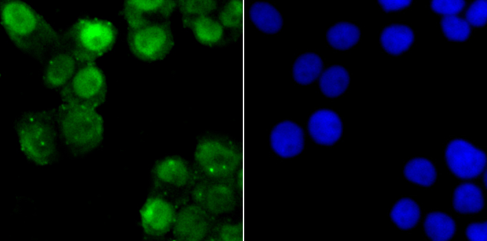
Fig3: ICC staining APE1 in LOVO cells (green). The nuclear counter stain is DAPI (blue). Cells were fixed in paraformaldehyde, permeabilised with 0.25% Triton X100/PBS.Application
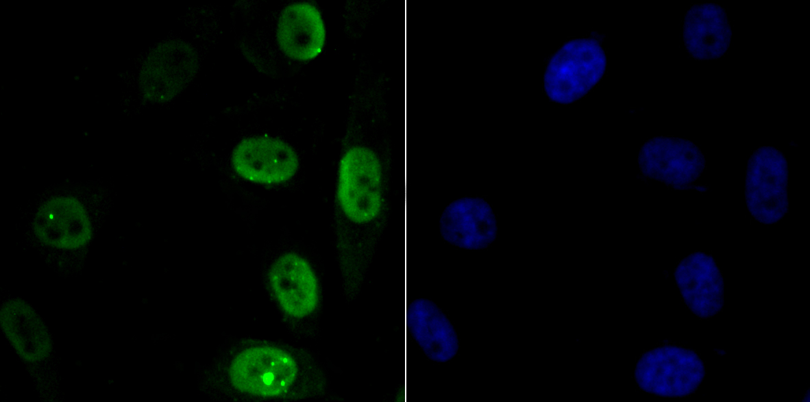
Fig4: ICC staining APE1 in PC-3M cells (green). The nuclear counter stain is DAPI (blue). Cells were fixed in paraformaldehyde, permeabilised with 0.25% Triton X100/PBS.Application
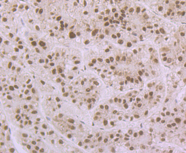
Fig5: Immunohistochemical analysis of paraffin-embedded human liver tissue using anti-APE1 antibody. Counter stained with hematoxylin.Application
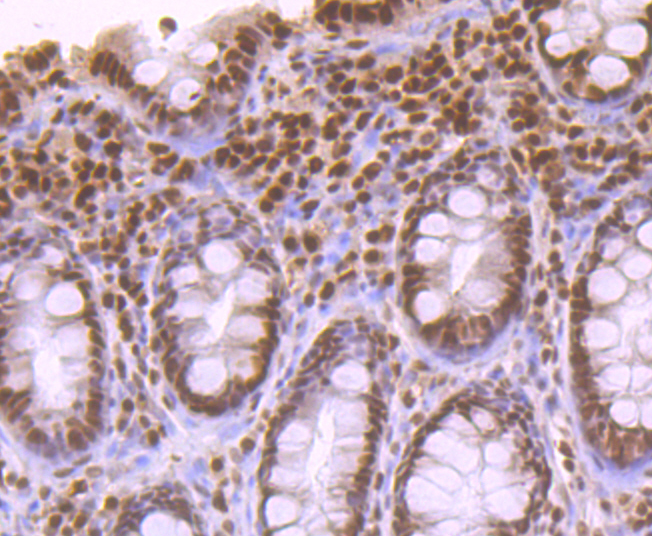
Fig6: Immunohistochemical analysis of paraffin-embedded human colon tissue using anti-APE1 antibody. Counter stained with hematoxylin.Application
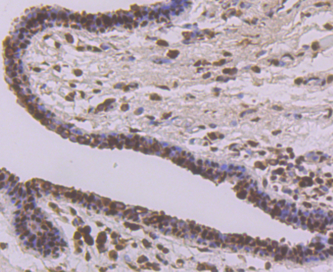
Fig7: Immunohistochemical analysis of paraffin-embedded human breast cancer tissue using anti-APE1 antibody. Counter stained with hematoxylin.Application
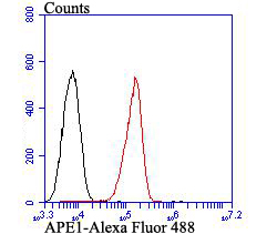
Fig8: Flow cytometric analysis of A549 cells with APE1 antibody at 1/100 dilution (red) compared with an unlabelled control (cells without incubation with primary antibody; black). Alexa Fluor 488-conjugated goat anti-rabbit IgG was used as the secondary antibody.| Product Name | APE1 |
|---|---|
| Antibody Type | Primary Antibodies |
| Antigen Alias | AP endonuclease 1 antibody AP endonuclease class I antibody AP lyase antibody APE 1 antibody APE antibody APE-1 antibody APEN antibody APEX 1 antibody APEX antibody APEX nuclease (multifunctional DNA repair enzyme) 1 antibody Apex nuclease 1 antibody APEX nuclease antibody APEX1 antibody APEX1_HUMAN antibody Apurinic endonuclease antibody Apurinic-apyrimidinic endonuclease 1 antibody Apurinic/apyrimidinic (abasic) endonuclease antibody Apurinic/apyrimidinic endonuclease 1 antibody Apurinic/apyrimidinic exonuclease antibody APX antibody BAP1 antibody Deoxyribonuclease (apurinic or apyrimidinic) antibody DNA (apurinic or apyrimidinic site) lyase antibody DNA-(apurinic or apyrimidinic site) lyase, mitochondrial antibody EC 4.2.99.18 antibody HAP 1 antibody HAP1 antibody Human Apurinic endonuclease 1 antibody MGC139790 antibody Multifunctional DNA repair enzyme antibody Redox factor 1 antibody Redox factor-1 antibody REF 1 antibody REF 1 protein antibody REF-1 antibody REF1 antibody REF1 protein antibody |
| Product description | The role of transcription factors in the regulation of gene expression is well established. Although the activity of these factors can be regulated by phosphorylation, evidence has indicated regulation of DNA binding mediated by changes in reduction-oxidation (redox) status. Mutational analysis has identified a single conserved cysteine residue mapping within the DNA binding domains of Fos and Jun. Chemical oxidation or modification of this cysteine residue inhibits the DNA binding activity of Fos and Jun. A similar mode of regulation has been recently proposed for other nuclear transcription factors. Oxidation is reversible by these compounds or by a cellular redox/DNA repair protein identified originally as Ref-1 (redox factor 1). Ref-1 is identical to a previously characterized DNA repair enzyme designated HAP1, APE or APEX. |
| Immunogen | Recombinant protein with Human APE1 aa 1-250. |
| Clonality | Polyclonal |
|---|---|
| Isotype | IgG |
| Host Species | Rabbit |
| Tested Applications | WBICCIHCFC |
| WB:1:500-1:2,000 ICC:1:500-1:1,000 IHC:1:100-1:500 FC:1:50-1:100 |
|
| Species Reactivity | HumanMouseRat |
| Concentration | 1mg/ml |
| Alternative Names | AP endonuclease 1 antibody
AP endonuclease class I antibody
AP lyase antibody
APE 1 antibody
APE antibody
APE-1 antibody
APEN antibody
APEX 1 antibody
APEX antibody
APEX nuclease (multifunctional DNA repair enzyme) 1 antibody
Apex nuclease 1 antibody
APEX nuclease antibody
APEX1 antibody
APEX1_HUMAN antibody
Apurinic endonuclease antibody
Apurinic-apyrimidinic endonuclease 1 antibody
Apurinic/apyrimidinic (abasic) endonuclease antibody
Apurinic/apyrimidinic endonuclease 1 antibody
Apurinic/apyrimidinic exonuclease antibody
APX antibody
BAP1 antibody
Deoxyribonuclease (apurinic or apyrimidinic) antibody
DNA (apurinic or apyrimidinic site) lyase antibody
DNA-(apurinic or apyrimidinic site) lyase mitochondrial antibody EC 4.2.99.18 antibody HAP 1 antibody HAP1 antibody Human Apurinic endonuclease 1 antibody MGC139790 antibody Multifunctional DNA repair enzyme antibody Redox factor 1 antibody Redox factor-1 antibody REF 1 antibody REF 1 protein antibody REF-1 antibody REF1 antibody REF1 protein antibody |
|---|---|
| Molecular Weight(MW) | 36 kDa |
| Cellular Localization | Nucleus. Endoplasmic reticulum. |
Application
Fig1: Western blot analysis of APE1 on different lysates using anti-APE1 antibody at 1/1,000 dilution. Positive control: Lane1: HL-60 Lane2: Human skin tissue Lane3: MCF-7 Lane4: Mouse placenta tissue
Application
Fig2: ICC staining APE1 in HepG2 cells (green). The nuclear counter stain is DAPI (blue). Cells were fixed in paraformaldehyde, permeabilised with 0.25% Triton X100/PBS.
Application
Fig3: ICC staining APE1 in LOVO cells (green). The nuclear counter stain is DAPI (blue). Cells were fixed in paraformaldehyde, permeabilised with 0.25% Triton X100/PBS.
Application
Fig4: ICC staining APE1 in PC-3M cells (green). The nuclear counter stain is DAPI (blue). Cells were fixed in paraformaldehyde, permeabilised with 0.25% Triton X100/PBS.
Application
Fig5: Immunohistochemical analysis of paraffin-embedded human liver tissue using anti-APE1 antibody. Counter stained with hematoxylin.
Application
Fig6: Immunohistochemical analysis of paraffin-embedded human colon tissue using anti-APE1 antibody. Counter stained with hematoxylin.
Application
Fig7: Immunohistochemical analysis of paraffin-embedded human breast cancer tissue using anti-APE1 antibody. Counter stained with hematoxylin.
Application
Fig8: Flow cytometric analysis of A549 cells with APE1 antibody at 1/100 dilution (red) compared with an unlabelled control (cells without incubation with primary antibody; black). Alexa Fluor 488-conjugated goat anti-rabbit IgG was used as the secondary antibody.| Positive Control | HL-60, human skin tissue lysate, mouse placenta tissue lysate, MCF-7, HepG2, LOVO, PC-3M, human liver tissue, human colon tissue, human breast cancer tissue, A549. |
|---|---|
| Application Notes | WB:1:500-1:2,000 ICC:1:500-1:1,000 IHC:1:100-1:500 FC:1:50-1:100 |
| Form | Liquid |
|---|---|
| Storage Instructions | Store at +4℃ after thawing. Aliquot store at -20℃. Avoid repeated freeze / thaw cycles. |
| Storage Buffer | 1*TBS (pH7.4), 0.5%BSA, 50%Glycerol. Preservative: 0.05% Sodium Azide. |


