Application
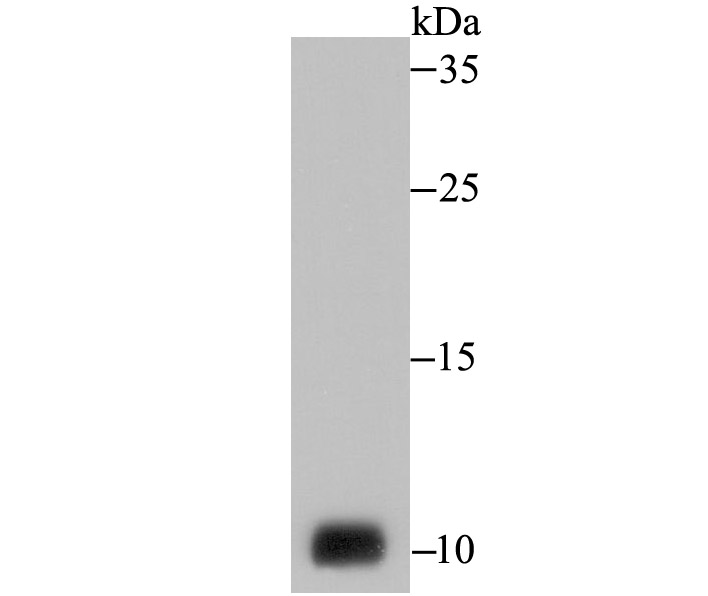
Fig1: Western blot analysis of NEDD8 on PC-3M cell lysate using anti-NEDD8 antibody at 1/1,000 dilution.Application
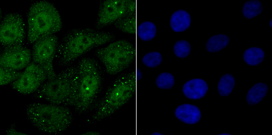
Fig2: ICC staining NEDD8 in HepG2 cells (green). The nuclear counter stain is DAPI (blue). Cells were fixed in paraformaldehyde, permeabilised with 0.25% Triton X100/PBS.Application
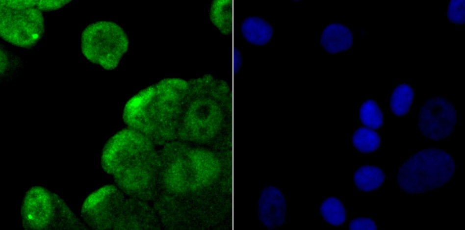
Fig3: ICC staining NEDD8 in MCF-7 cells (green). The nuclear counter stain is DAPI (blue). Cells were fixed in paraformaldehyde, permeabilised with 0.25% Triton X100/PBS.Application
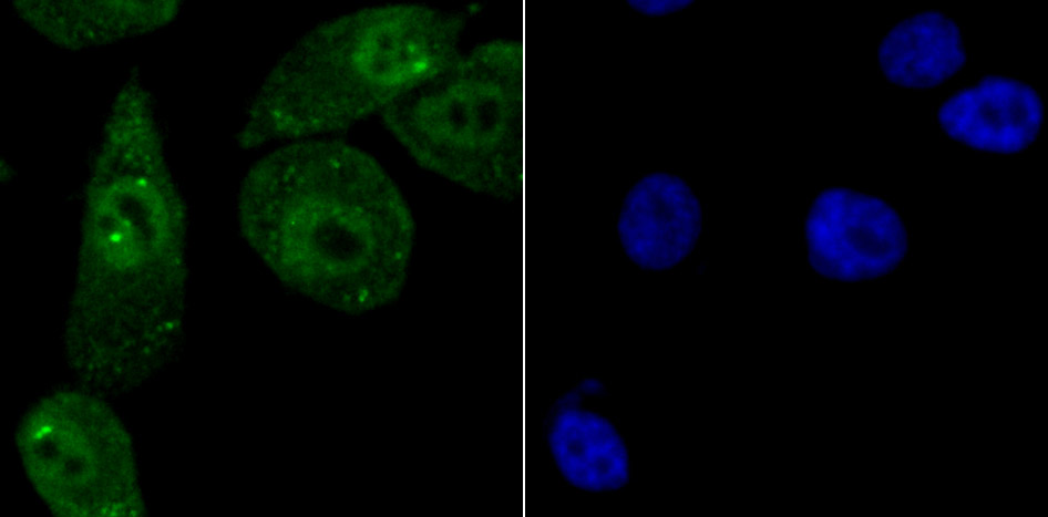
Fig4: ICC staining NEDD8 in PC-3M cells (green). The nuclear counter stain is DAPI (blue). Cells were fixed in paraformaldehyde, permeabilised with 0.25% Triton X100/PBS.Application
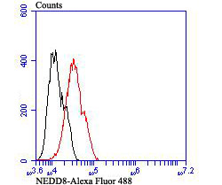
Fig5: Flow cytometric analysis of MCF-7 cells with NEDD8 antibody at 1/100 dilution (red) compared with an unlabelled control (cells without incubation with primary antibody; black). Alexa Fluor 488-conjugated goat anti-rabbit IgG was used as the secondary antibody.| Product Name | NEDD8 |
|---|---|
| Antibody Type | Primary Antibodies |
| Antigen Alias | FLJ43224 antibody MGC104393 antibody MGC125896 antibody MGC125897 antibody NED8 antibody NEDD 8 antibody NEDD-8 antibody Nedd8 antibody NEDD8_HUMAN antibody Neddylin antibody Neural precursor cell expressed developmentally down regulated 8 antibody Neural precursor cell expressed developmentally down regulated gene 8 antibody Neural precursor cell expressed developmentally down-regulated protein 8 antibody Rub1 antibody Ubiquitin like protein Nedd 8 antibody Ubiquitin like protein Nedd8 antibody Ubiquitin-like protein Nedd8 antibody |
| Product description | NEDD8 is a ubiquitin (Ub)-like molecule that shares 80% homology with ubiquitin, a protein known to modify and target various proteins for proteolytic degradation. NEDD8 and the corresponding yeast homolog Rub1 are activated by the E1 ubiquitin activating enzyme UBA2, which forms isopeptide linkages between thio esters. Similar to the Ub-mediated proteolytic pathway, NEDD8 is covalently coupled to an E3 Ub ligase by the E2 conjugating enzyme, Ubc12. NEDD8 preferentially associates with the CUL-2 protein in the E3 ligase complex CBCVHL, which consists of cullin proteins associating with elongin B/C and the tumor suppressor F-box protein, von Hippel Lindau (VHL). NEDD8 is predominantly localized to the nucleus and is highly expressed in adult heart and skeletal muscle. In vitro studies indicate that NEDD8 tetramers are also able to bind to the 26S Proteasome, and that they assemble into complexes with conserved Ub-like moieties, suggesting that NEDD8 may regulate proteolysis of intracellular proteins similar to other Ub-mediated pathways. |
| Immunogen | Recombinant protein |
| Clonality | Polyclonal |
|---|---|
| Isotype | IgG |
| Host Species | Rabbit |
| Tested Applications | WBICCFC |
| WB:1:500-1:1,000 ICC:1:50-1:100 FC:1:50-1:100 |
|
| Species Reactivity | Human |
| Concentration | 1mg/ml |
| Alternative Names | FLJ43224 antibody MGC104393 antibody MGC125896 antibody MGC125897 antibody NED8 antibody NEDD 8 antibody NEDD-8 antibody Nedd8 antibody NEDD8_HUMAN antibody Neddylin antibody Neural precursor cell expressed developmentally down regulated 8 antibody Neural precursor cell expressed developmentally down regulated gene 8 antibody Neural precursor cell expressed developmentally down-regulated protein 8 antibody Rub1 antibody Ubiquitin like protein Nedd 8 antibody Ubiquitin like protein Nedd8 antibody Ubiquitin-like protein Nedd8 antibody |
|---|---|
| Molecular Weight(MW) | 9 kDa |
| Cellular Localization | Nucleus. |
| SwissProt ID | Q15843 |
|---|
Application
Fig1: Western blot analysis of NEDD8 on PC-3M cell lysate using anti-NEDD8 antibody at 1/1,000 dilution.
Application
Fig2: ICC staining NEDD8 in HepG2 cells (green). The nuclear counter stain is DAPI (blue). Cells were fixed in paraformaldehyde, permeabilised with 0.25% Triton X100/PBS.
Application
Fig3: ICC staining NEDD8 in MCF-7 cells (green). The nuclear counter stain is DAPI (blue). Cells were fixed in paraformaldehyde, permeabilised with 0.25% Triton X100/PBS.
Application
Fig4: ICC staining NEDD8 in PC-3M cells (green). The nuclear counter stain is DAPI (blue). Cells were fixed in paraformaldehyde, permeabilised with 0.25% Triton X100/PBS.
Application
Fig5: Flow cytometric analysis of MCF-7 cells with NEDD8 antibody at 1/100 dilution (red) compared with an unlabelled control (cells without incubation with primary antibody; black). Alexa Fluor 488-conjugated goat anti-rabbit IgG was used as the secondary antibody.| Positive Control | PC-3M, HepG2, MCF-7. |
|---|---|
| Application Notes | WB:1:500-1:1,000 ICC:1:50-1:100 FC:1:50-1:100 |
| Form | Liquid |
|---|---|
| Storage Instructions | Store at +4℃ after thawing. Aliquot store at -20℃ or -80℃. Avoid repeated freeze / thaw cycles. |
| Storage Buffer | 1*TBS (pH7.4), 0.5%BSA, 50%Glycerol. Preservative: 0.05% Sodium Azide. |


