Application
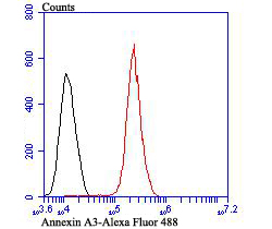
Fig6: Flow cytometric analysis of A431 cells with Annexin A3 antibody at 1/100 dilution (red) compared with an unlabelled control (cells without incubation with primary antibody; black). Alexa Fluor 488-conjugated goat anti-rabbit IgG was used as the secondary antibody.Application
Fig6: Flow cytometric analysis of A431 cells with Annexin A3 antibody at 1/100 dilution (red) compared with an unlabelled control (cells without incubation with primary antibody; black). Alexa Fluor 488-conjugated goat anti-rabbit IgG was used as the secondary antibody.Application
Fig6: Flow cytometric analysis of A431 cells with Annexin A3 antibody at 1/100 dilution (red) compared with an unlabelled control (cells without incubation with primary antibody; black). Alexa Fluor 488-conjugated goat anti-rabbit IgG was used as the secondary antibody.Application
Fig6: Flow cytometric analysis of A431 cells with Annexin A3 antibody at 1/100 dilution (red) compared with an unlabelled control (cells without incubation with primary antibody; black). Alexa Fluor 488-conjugated goat anti-rabbit IgG was used as the secondary antibody.Application
Fig6: Flow cytometric analysis of A431 cells with Annexin A3 antibody at 1/100 dilution (red) compared with an unlabelled control (cells without incubation with primary antibody; black). Alexa Fluor 488-conjugated goat anti-rabbit IgG was used as the secondary antibody.Application
Fig6: Flow cytometric analysis of A431 cells with Annexin A3 antibody at 1/100 dilution (red) compared with an unlabelled control (cells without incubation with primary antibody; black). Alexa Fluor 488-conjugated goat anti-rabbit IgG was used as the secondary antibody.| Product Name | Annexin A3 |
|---|---|
| Antibody Type | Primary Antibodies |
| Antigen Alias | 2-cyclic phosphate 2-phosphohydrolase antibody 35-alpha calcimedin antibody Annexin A3 antibody Annexin III antibody Annexin-3 antibody ANX3 antibody ANXA 3 antibody Anxa3 antibody ANXA3_HUMAN antibody Calcimedin 35 alpha antibody Inositol 1 2 cyclic phosphate 2 phosphohydrolase antibody Inositol 1 antibody Lipocortin III antibody OTTHUMP00000160701 antibody OTTHUMP00000219237 antibody OTTHUMP00000219238 antibody PAP III antibody PAP-III antibody Placental anticoagulant protein III antibody |
| Product description | The annexin family of calcium-binding proteins is composed of at least ten mammalian genes and is characterized by a conserved core domain, which binds phospholipids in a Ca2+-dependent manner, and a unique amino-terminal region, which may confer binding specificity. Annexin family members have been implicated as regulators of such diverse processes as ion flux, endocytosis, exocytosis and cellular adhesion. Annexin III, also known as ANXA3, Annexin A3, Lipocortin III, PAP-III (placental anticoagulant protein III), 35-α calcimedin or ANX3, belongs to the annexin family and contains four Annexin repeats. Annexin III exhibits a variety of functions, including anti-coagulent properties, an inhibitory role towards phospholipase A2 (PLA2) and an enzymatic function, cleaving the cyclic bond of inositol 1,2-cyclic phosphate to produce inositol 1-phosphate. |
| Immunogen | Recombinant protein with Human Annexin A3 aa 1-350. |
| Clonality | Polyclonal |
|---|---|
| Isotype | IgG |
| Host Species | Rabbit |
| Tested Applications | WBIHCFC |
| WB:1:500-1:2,000 IHC:1:50-1:200 FC:1:50-1:100 |
|
| Species Reactivity | HumanMouseRat |
| Concentration | 1mg/ml |
| Alternative Names | 2-cyclic phosphate 2-phosphohydrolase antibody 35-alpha calcimedin antibody Annexin A3 antibody Annexin III antibody Annexin-3 antibody ANX3 antibody ANXA 3 antibody Anxa3 antibody ANXA3_HUMAN antibody Calcimedin 35 alpha antibody Inositol 1 2 cyclic phosphate 2 phosphohydrolase antibody Inositol 1 antibody Lipocortin III antibody OTTHUMP00000160701 antibody OTTHUMP00000219237 antibody OTTHUMP00000219238 antibody PAP III antibody PAP-III antibody Placental anticoagulant protein III antibody |
|---|---|
| Molecular Weight(MW) | 36 kDa |
| Cellular Localization | Cytoplasm. Secreted. Plasma Membrane. |
Application
Fig6: Flow cytometric analysis of A431 cells with Annexin A3 antibody at 1/100 dilution (red) compared with an unlabelled control (cells without incubation with primary antibody; black). Alexa Fluor 488-conjugated goat anti-rabbit IgG was used as the secondary antibody.
Application
Fig6: Flow cytometric analysis of A431 cells with Annexin A3 antibody at 1/100 dilution (red) compared with an unlabelled control (cells without incubation with primary antibody; black). Alexa Fluor 488-conjugated goat anti-rabbit IgG was used as the secondary antibody.
Application
Fig6: Flow cytometric analysis of A431 cells with Annexin A3 antibody at 1/100 dilution (red) compared with an unlabelled control (cells without incubation with primary antibody; black). Alexa Fluor 488-conjugated goat anti-rabbit IgG was used as the secondary antibody.
Application
Fig6: Flow cytometric analysis of A431 cells with Annexin A3 antibody at 1/100 dilution (red) compared with an unlabelled control (cells without incubation with primary antibody; black). Alexa Fluor 488-conjugated goat anti-rabbit IgG was used as the secondary antibody.
Application
Fig6: Flow cytometric analysis of A431 cells with Annexin A3 antibody at 1/100 dilution (red) compared with an unlabelled control (cells without incubation with primary antibody; black). Alexa Fluor 488-conjugated goat anti-rabbit IgG was used as the secondary antibody.
Application
Fig6: Flow cytometric analysis of A431 cells with Annexin A3 antibody at 1/100 dilution (red) compared with an unlabelled control (cells without incubation with primary antibody; black). Alexa Fluor 488-conjugated goat anti-rabbit IgG was used as the secondary antibody.| Positive Control | Siha, Hela, rat spleen tissue lysate, rat liver tissue lysate, mouse lung tissue lysate, rat kidney tissue, human colon cancer tissue, human spleen tissue, human prostate tissue, A431. |
|---|---|
| Application Notes | WB:1:500-1:2,000 IHC:1:50-1:200 FC:1:50-1:100 |
| Form | Liquid |
|---|---|
| Storage Instructions | Store at +4℃ after thawing. Aliquot store at -20℃. Avoid repeated freeze / thaw cycles. |
| Storage Buffer | 1*TBS (pH7.4), 0.5%BSA, 50%Glycerol. Preservative: 0.05% Sodium Azide. |


